Sepsis
Sepsis is a spectrum of disease, where there is a systemic and dysregulated host response to an infection.[65]Singer M, Deutschman CS, Seymour CW, et al. The Third International Consensus Definitions for sepsis and septic shock (Sepsis-3). JAMA. 2016 Feb 23;315(8):801-10.
https://www.doi.org/10.1001/jama.2016.0287
http://www.ncbi.nlm.nih.gov/pubmed/26903338?tool=bestpractice.com
Presentation ranges from subtle, non-specific symptoms (e.g., feeling unwell with a normal temperature) to severe symptoms with evidence of multi-organ dysfunction and septic shock. Patients may have signs of tachycardia, tachypnoea, hypotension, fever or hypothermia, poor capillary refill, mottled or ashen skin, cyanosis, newly altered mental state or reduced urine output.[66]National Institute for Health and Care Excellence. Suspected sepsis: recognition, diagnosis and early management. Mar 2024 [internet publication].
https://www.nice.org.uk/guidance/ng51
Sepsis and septic shock are medical emergencies.
Risk factors for sepsis in children include: age under 1 year, impaired immunity (due to illness or drugs), recent surgery or other invasive procedures, any breach of skin integrity (e.g., cuts, burns), and indwelling lines or catheters.[66]National Institute for Health and Care Excellence. Suspected sepsis: recognition, diagnosis and early management. Mar 2024 [internet publication].
https://www.nice.org.uk/guidance/ng51
Early recognition of sepsis is essential because early treatment improves outcomes.[66]National Institute for Health and Care Excellence. Suspected sepsis: recognition, diagnosis and early management. Mar 2024 [internet publication].
https://www.nice.org.uk/guidance/ng51
[67]Weiss SL, Peters MJ, Alhazzani W, et al. Surviving sepsis campaign international guidelines for the management of septic shock and sepsis-associated organ dysfunction in children. Intensive Care Med. 2020 Feb;46(suppl 1):10-67.
https://link.springer.com/article/10.1007%2Fs00134-019-05878-6
http://www.ncbi.nlm.nih.gov/pubmed/32030529?tool=bestpractice.com
[Evidence C]4fbddd42-78e2-41c0-9f27-d6fdae81a072guidelineCWhat are the effects of early versus late initiation of empiric antimicrobial treatment in children with or at risk of developing sepsis or severe sepsis?[66]National Institute for Health and Care Excellence. Suspected sepsis: recognition, diagnosis and early management. Mar 2024 [internet publication].
https://www.nice.org.uk/guidance/ng51
However, detection can be challenging because the clinical presentation of sepsis can be subtle and non-specific. A low threshold for suspecting sepsis is therefore important.
The key to early recognition is the systematic identification of any patient who has signs or symptoms suggestive of infection and is at risk of deterioration due to organ dysfunction. Criteria to identify sepsis and septic shock in children and young people under the age of 18 years have been developed.[68]Schlapbach LJ, Watson RS, Sorce LR, et al. International consensus criteria for pediatric sepsis and septic shock. JAMA. 2024 Feb 27;331(8):665-74.
https://jamanetwork.com/journals/jama/fullarticle/2814297
http://www.ncbi.nlm.nih.gov/pubmed/38245889?tool=bestpractice.com
Several other risk stratification approaches exist. All rely on a structured clinical assessment and recording of the patient's vital signs.[66]National Institute for Health and Care Excellence. Suspected sepsis: recognition, diagnosis and early management. Mar 2024 [internet publication].
https://www.nice.org.uk/guidance/ng51
[67]Weiss SL, Peters MJ, Alhazzani W, et al. Surviving sepsis campaign international guidelines for the management of septic shock and sepsis-associated organ dysfunction in children. Intensive Care Med. 2020 Feb;46(suppl 1):10-67.
https://link.springer.com/article/10.1007%2Fs00134-019-05878-6
http://www.ncbi.nlm.nih.gov/pubmed/32030529?tool=bestpractice.com
[68]Schlapbach LJ, Watson RS, Sorce LR, et al. International consensus criteria for pediatric sepsis and septic shock. JAMA. 2024 Feb 27;331(8):665-74.
https://jamanetwork.com/journals/jama/fullarticle/2814297
http://www.ncbi.nlm.nih.gov/pubmed/38245889?tool=bestpractice.com
[69]Royal College of Physicians. National Early Warning Score (NEWS) 2. Dec 2017 [internet publication].
https://www.rcplondon.ac.uk/projects/outputs/national-early-warning-score-news-2
[70]American College of Emergency Physicians (ACEP) Expert Panel on Sepsis. DART: an evidence-driven tool to guide the early recognition and treatment of sepsis and septic shock [internet publication].
https://poctools.acep.org/POCTool/Sepsis(DART)/276ed0a9-f24d-45f1-8d0c-e908a2758e5a
[71]Academy of Medical Royal Colleges. Statement on the initial antimicrobial treatment of sepsis. Oct 2022 [internet publication].
https://www.aomrc.org.uk/reports-guidance/statement-on-the-initial-antimicrobial-treatment-of-sepsis-v2-0
It is important to check local guidance for information on which approach your institution recommends. The timeline of ensuing investigations and treatment should be guided by this early assessment.[71]Academy of Medical Royal Colleges. Statement on the initial antimicrobial treatment of sepsis. Oct 2022 [internet publication].
https://www.aomrc.org.uk/reports-guidance/statement-on-the-initial-antimicrobial-treatment-of-sepsis-v2-0
Treatment guidelines have been produced by the Surviving Sepsis Campaign and remain the most widely accepted standards.[67]Weiss SL, Peters MJ, Alhazzani W, et al. Surviving sepsis campaign international guidelines for the management of septic shock and sepsis-associated organ dysfunction in children. Intensive Care Med. 2020 Feb;46(suppl 1):10-67.
https://link.springer.com/article/10.1007%2Fs00134-019-05878-6
http://www.ncbi.nlm.nih.gov/pubmed/32030529?tool=bestpractice.com
Within the first hour:[67]Weiss SL, Peters MJ, Alhazzani W, et al. Surviving sepsis campaign international guidelines for the management of septic shock and sepsis-associated organ dysfunction in children. Intensive Care Med. 2020 Feb;46(suppl 1):10-67.
https://link.springer.com/article/10.1007%2Fs00134-019-05878-6
http://www.ncbi.nlm.nih.gov/pubmed/32030529?tool=bestpractice.com
Follow institutional protocols for management of sepsis/septic shock in children; these improve the speed and reliability of care
Obtain blood cultures before administering antibiotics (provided this does not substantially delay antibiotic administration)
Administer broad-spectrum antibiotics
Administer crystalloid fluids, titrated to clinical signs of cardiac output and stopped if there is evidence of volume overload. Consult local protocols.
Use trends in blood lactate levels to guide resuscitation. If the child's hypotension is refractory to fluid resuscitation, consider use of vasopressors.[67]Weiss SL, Peters MJ, Alhazzani W, et al. Surviving sepsis campaign international guidelines for the management of septic shock and sepsis-associated organ dysfunction in children. Intensive Care Med. 2020 Feb;46(suppl 1):10-67.
https://link.springer.com/article/10.1007%2Fs00134-019-05878-6
http://www.ncbi.nlm.nih.gov/pubmed/32030529?tool=bestpractice.com
For more information on sepsis, please see our topic Sepsis in children.
Anaphylaxis
Patients may present with airway compromise, difficulty breathing, hypotension, and/or tachycardia and collapse. Emergency intervention includes calling for help, giving intramuscular adrenaline (epinephrine), oxygen, and supportive measures.[72]Shaker MS, Wallace DV, Golden DBK, et al. Anaphylaxis-a 2020 practice parameter update, systematic review, and Grading of Recommendations, Assessment, Development and Evaluation (GRADE) analysis. J Allergy Clin Immunol. 2020 Apr;145(4):1082-123.
https://www.jacionline.org/article/S0091-6749(20)30105-6/fulltext
http://www.ncbi.nlm.nih.gov/pubmed/32001253?tool=bestpractice.com
[73]Golden DBK, Wang J, Waserman S, et al. Anaphylaxis: a 2023 practice parameter update. Ann Allergy Asthma Immunol. 2024 Feb;132(2):124-76.
https://www.annallergy.org/article/S1081-1206(23)01304-2/fulltext
http://www.ncbi.nlm.nih.gov/pubmed/38108678?tool=bestpractice.com
Toxic epidermal necrolysis (TEN) and Stevens-Johnson syndrome (SJS)
TEN and SJS constitute a spectrum of severe generalised exfoliative dermatitis with associated involvement of ≥2 mucosal surfaces (oral, conjunctival, anogenital).[43]Miliszewski MA, Kirchhof MG, Sikora S, et al. Stevens-Johnson syndrome and toxic epidermal necrolysis: an analysis of triggers and implications for improving prevention. Am J Med. 2016 Nov;129(11):1221-5.
http://www.ncbi.nlm.nih.gov/pubmed/27086495?tool=bestpractice.com
Skin lesions may be initially targetoid (with no central blistering), although they often become confluent and bullous. Nikolsky's sign (blister induced with lateral pressure) may be noted within affected areas.[43]Miliszewski MA, Kirchhof MG, Sikora S, et al. Stevens-Johnson syndrome and toxic epidermal necrolysis: an analysis of triggers and implications for improving prevention. Am J Med. 2016 Nov;129(11):1221-5.
http://www.ncbi.nlm.nih.gov/pubmed/27086495?tool=bestpractice.com
The lesions are painful and the patient appears acutely unwell. [Figure caption and citation for the preceding image starts]: Stevens-Johnson syndrome: targetoid lesions and epidermal lossFrom the personal collection of Dr A. Kowal-Vern [Citation ends].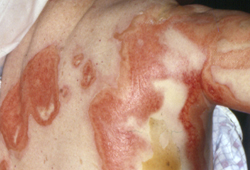 [Figure caption and citation for the preceding image starts]: Stevens-Johnson syndrome: epidermal loss on soles of feetFrom the personal collection of Dr A. Kowal-Vern [Citation ends].
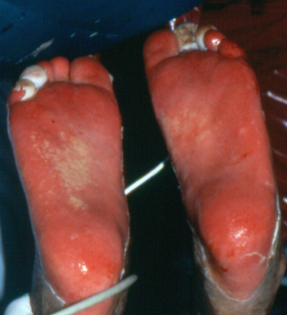 [Figure caption and citation for the preceding image starts]: Toxic epidermal necrolysis with epidermal loss, ocular involvement, and ecthyma gangrenosumFrom the personal collection of Dr A. Kowal-Vern [Citation ends].
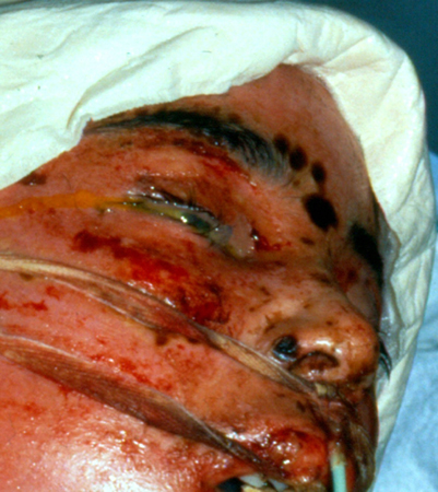
Initial management includes:
Drug reaction with eosinophilia and systemic symptoms (DRESS) syndrome
The presentation of DRESS syndrome resembles a morbilliform drug eruption, but the patient is more unwell, often with fever, abdominal pain, lymphadenopathy, and facial swelling.[45]Husain Z, Reddy BY, Schwartz RA. DRESS syndrome: part I. Clinical perspectives. J Am Acad Dermatol. 2013 May;68(5):693.e1-14; quiz 706-8.
http://www.ncbi.nlm.nih.gov/pubmed/23602182?tool=bestpractice.com
[46]Adwan MH. Drug reaction with eosinophilia and systemic symptoms (DRESS) syndrome and the rheumatologist. Curr Rheumatol Rep. 2017 Jan;19(1):3.
http://www.ncbi.nlm.nih.gov/pubmed/28138822?tool=bestpractice.com
The time interval between intake of the offending medication and symptoms also tends to be longer, often 2 to 6 weeks. Typically, the rash starts on the face, upper torso, and upper extremities, then spreads to the lower extremities. Liver involvement is common, presenting with hepatosplenomegaly and elevated transaminases. Full blood count (FBC) may show marked eosinophilia and leukocytosis.[45]Husain Z, Reddy BY, Schwartz RA. DRESS syndrome: part I. Clinical perspectives. J Am Acad Dermatol. 2013 May;68(5):693.e1-14; quiz 706-8.
http://www.ncbi.nlm.nih.gov/pubmed/23602182?tool=bestpractice.com
Immediate withdrawal of the medication is indicated. Treatment with oral corticosteroids may be required. In severe hypersensitivity reactions that do not respond to corticosteroids, intravenous immunoglobulin (IVIG) can be tried.
Meningococcal disease
Meningococcal disease may progress rapidly, and clinical deterioration may continue despite the prompt institution of antimicrobial therapy. Initial assessment should follow the principles of paediatric advanced life support, with assessment of the patient's airway, breathing, and circulatory status, and establishment of secure large-calibre intravenous or intraosseous catheters for administration of fluids.[67]Weiss SL, Peters MJ, Alhazzani W, et al. Surviving sepsis campaign international guidelines for the management of septic shock and sepsis-associated organ dysfunction in children. Intensive Care Med. 2020 Feb;46(suppl 1):10-67.
https://link.springer.com/article/10.1007%2Fs00134-019-05878-6
http://www.ncbi.nlm.nih.gov/pubmed/32030529?tool=bestpractice.com
[Figure caption and citation for the preceding image starts]: Petechial rash in invasive meningococcal diseaseThomas AE, et al. BMJ. 2016 Mar 22;352:i1285 [Citation ends].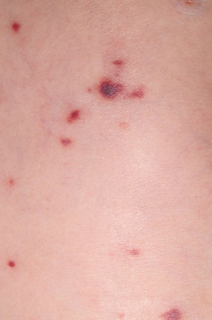
Diagnostic testing includes a blood culture and lumbar puncture (if it is safe to do so) for examination of cerebrospinal fluid in patients with suspected bacterial meningitis.[74]Heckenberg SG, Brouwer MC, van de Beek D, et al. Bacterial meningitis. Handb Clin Neurol. 2014;121:1361-75.
http://www.ncbi.nlm.nih.gov/pubmed/24365425?tool=bestpractice.com
Before performing a lumbar puncture, treat and stabilise any of the following: unprotected airway, respiratory compromise, shock, uncontrolled seizures, bleeding risk.[75]National Institute for Health and Care Excellence. Meningitis (bacterial) and meningococcal disease: recognition, diagnosis and management. Mar 2024 [internet publication].
https://www.nice.org.uk/guidance/ng240
Avoid lumbar puncture if the patient has extensive or rapidly evolving purpuric rash, risk factors for an evolving space-occupying lesion, clinical signs of raised intracranial pressure (reduced or fluctuating level of consciousness, relative bradycardia and hypertension, new focal neurological signs, papilloedema, or abnormal posturing, abnormal pupillary responses, or eye movements), or local infection at the site for lumbar puncture.[75]National Institute for Health and Care Excellence. Meningitis (bacterial) and meningococcal disease: recognition, diagnosis and management. Mar 2024 [internet publication].
https://www.nice.org.uk/guidance/ng240
Empirical antibiotics should be given as soon as possible to any child with suspected meningococcal disease.[67]Weiss SL, Peters MJ, Alhazzani W, et al. Surviving sepsis campaign international guidelines for the management of septic shock and sepsis-associated organ dysfunction in children. Intensive Care Med. 2020 Feb;46(suppl 1):10-67.
https://link.springer.com/article/10.1007%2Fs00134-019-05878-6
http://www.ncbi.nlm.nih.gov/pubmed/32030529?tool=bestpractice.com
Antibiotics should not be delayed if diagnostic assessment cannot be completed promptly.[75]National Institute for Health and Care Excellence. Meningitis (bacterial) and meningococcal disease: recognition, diagnosis and management. Mar 2024 [internet publication].
https://www.nice.org.uk/guidance/ng240
Acute lymphocytic leukaemia
Acute lymphocytic leukaemia is the most likely haematological cancer to present with cutaneous manifestations. Initially patients may require hydration (with diuresis and urinary alkalinisation) and allopurinol or rasburicase to prevent tumour lysis syndrome associated with hyperuricaemia. Transfusion of blood products, including platelet transfusion, may be required depending on symptoms and blood count.
Staphylococcal scalded skin syndrome (SSSS), toxic shock syndrome (TSS) and scarlet fever
SSSS is more likely in children and presents with moderate fever, generalised erythema, and blisters and erosions accentuated in the intertriginous areas. The diagnosis is primarily clinical, as blood and fluid cultures are generally negative. Treatment for SSSS is primarily supportive care (with fluid rehydration and topical wound care) and parenteral antibiotics to cover Staphylococcus aureus.
TSS is best managed with parenteral antibiotics targeting the causative organism (e.g., methicillin-sensitive or methicillin-resistant S aureus, Streptococcus pyogenes) and intensive supportive therapy. Mortality for TSS in children is approximately 2%.[55]Strom MA, Hsu DY, Silverberg JI. Prevalence, comorbidities and mortality of toxic shock syndrome in children and adults in the USA. Microbiol Immunol. 2017 Nov;61(11):463-73.
https://onlinelibrary.wiley.com/doi/full/10.1111/1348-0421.12539
http://www.ncbi.nlm.nih.gov/pubmed/28892185?tool=bestpractice.com
Approximately 90% of scarlet fever cases occur in children under 10 years old.[76]UK Health Security Agency. Scarlet fever: symptoms, diagnosis and treatment. Mar 2019 [internet publication].
https://www.gov.uk/government/publications/scarlet-fever-symptoms-diagnosis-treatment/scarlet-fever-factsheet
It is usually a mild illness, but is highly infectious. It presents with a generalised, erythematous rash, which feels like sandpaper and is often preceded by a sore throat (pharyngitis, tonsillitis). Pharyngeal erythema with exudates, palatal petechiae, and a red, swollen (strawberry) tongue are suggestive features. Clinicians are advised to maintain a high index of suspicion, as early recognition and prompt initiation of specific and supportive therapy for patients with invasive group A streptococcal infection can be life-saving. Prompt treatment of scarlet fever with antibiotics is recommended to reduce risk of possible complications, including invasive group A streptococcus, and to limit onward transmission.
If a rapid antigen detection test (RADT) for group A streptococcus is not available, antibiotics can be prescribed subsequent to a clinical diagnosis of scarlet fever by a health professional.[77]UK Health Security Agency. Scarlet fever: managing outbreaks in schools and nurseries. Apr 2023 [internet publication].
https://www.gov.uk/government/publications/scarlet-fever-managing-outbreaks-in-schools-and-nurseries
In countries where RADTs for scarlet fever are available, a positive test result may be required before starting antibiotics (patients with clear viral symptoms don't need testing for group A streptococcal bacteria).[78]US Centers for Disease Control and Prevention. Clinical guidance for Group A streptococcal pharyngitis. Mar 2024 [internet publication].
https://www.cdc.gov/group-a-strep/hcp/clinical-guidance/strep-throat.html
If there is uncertainty about the diagnosis, obtain a throat swab prior to commencing antibiotics.[78]US Centers for Disease Control and Prevention. Clinical guidance for Group A streptococcal pharyngitis. Mar 2024 [internet publication].
https://www.cdc.gov/group-a-strep/hcp/clinical-guidance/strep-throat.html
[79]UK Health Security Agency. Group A streptococcal infections: activity during the 2022 to 2023 season. Jun 2023 [internet publication].
https://www.gov.uk/government/publications/group-a-streptococcal-infections-activity-during-the-2022-to-2023-season
[80]Shulman ST, Bisno AL, Clegg HW, et al. Clinical practice guideline for the diagnosis and management of group A streptococcal pharyngitis: 2012 update by the Infectious Diseases Society of America. Clin Infect Dis. 2012 Nov 15;55(10):e86-102.
https://www.ncbi.nlm.nih.gov/pmc/articles/PMC7108032
http://www.ncbi.nlm.nih.gov/pubmed/22965026?tool=bestpractice.com
[81]Gerber MA, Baltimore RS, Eaton CB, et al. Prevention of rheumatic fever and diagnosis and treatment of acute streptococcal pharyngitis: a scientific statement from the American Heart Association Rheumatic Fever, Endocarditis, and Kawasaki Disease Committee of the Council on Cardiovascular Disease in the Young, the Interdisciplinary Council on Functional Genomics and Translational Biology, and the Interdisciplinary Council on Quality of Care and Outcomes Research: endorsed by the American Academy of Pediatrics. Circulation. 2009 Mar 24;119(11):1541-51.
https://www.ahajournals.org/doi/10.1161/CIRCULATIONAHA.109.191959?url_ver=Z39.88-2003&rfr_id=ori:rid:crossref.org&rfr_dat=cr_pub%20%200pubmed
http://www.ncbi.nlm.nih.gov/pubmed/19246689?tool=bestpractice.com
Treatment of scarlet fever with antibiotics based on clinical diagnosis alone should follow in-country clinical guidelines.
According to the UK Health Security Agency, notifications of scarlet fever and invasive GAS (iGAS) disease in England were higher than expected from September 2022 to February 2023, with the peak observed in December 2022.[79]UK Health Security Agency. Group A streptococcal infections: activity during the 2022 to 2023 season. Jun 2023 [internet publication].
https://www.gov.uk/government/publications/group-a-streptococcal-infections-activity-during-the-2022-to-2023-season
[82]UK Health Security Agency. UKHSA update on scarlet fever and invasive group A strep. May 2023 [internet publication].
https://www.gov.uk/government/news/ukhsa-update-on-scarlet-fever-and-invasive-group-a-strep-1
The notifications have significantly reduced since then and are now in line with the expected number for the time of year.[79]UK Health Security Agency. Group A streptococcal infections: activity during the 2022 to 2023 season. Jun 2023 [internet publication].
https://www.gov.uk/government/publications/group-a-streptococcal-infections-activity-during-the-2022-to-2023-season
Other countries experiencing an increased incidence of scarlet fever and iGAS disease during this period include France, Ireland, the Netherlands, and Sweden. The increase was particularly marked during the second half of 2022.[83]World Health Organization. Increased incidence of scarlet fever and invasive group A streptococcus infection - multi-country. Dec 2022 [internet publication].
https://www.who.int/emergencies/disease-outbreak-news/item/2022-DON429
Infective endocarditis
If the child with suspected endocarditis presents with significant upset (e.g., headache, meningeal signs, dyspnoea on exertion, or stroke symptoms), urgent assessment and treatment with empirical broad-spectrum antibiotic therapy is required.[84]Dixon G, Christov G. Infective endocarditis in children: an update. Curr Opin Infect Dis. 2017 Jun;30(3):257-67.
http://www.ncbi.nlm.nih.gov/pubmed/28319472?tool=bestpractice.com
Initial laboratory studies should include FBC and electrolyte panel, with blood cultures and urine for urinalysis taken before antibiotic administration if possible. An ECG should be obtained.[85]Webb R, Voss L, Roberts S, et al. Infective endocarditis in New Zealand children 1994-2012. Pediatr Infect Dis J. 2014 May;33(5):437-42.
http://www.ncbi.nlm.nih.gov/pubmed/24378941?tool=bestpractice.com
Fungal endocarditis is unusual in children, but can be seen with indwelling central catheters, which may need to be removed urgently.[52]Baltimore RS, Gewitz M, Baddour LM, et al; American Heart Association Rheumatic Fever, Endocarditis, and Kawasaki Disease Committee of the Council on Cardiovascular Disease in the Young and the Council on Cardiovascular and Stroke Nursing. Infective endocarditis in childhood: 2015 update: a scientific statement from the American Heart Association. Circulation. 2015 Oct 13;132(15):1487-515.
https://www.ahajournals.org/doi/full/10.1161/cir.0000000000000298
http://www.ncbi.nlm.nih.gov/pubmed/26373317?tool=bestpractice.com
Rheumatic fever
Most patients present with fever and a history or evidence of a recent group A streptococcal infection, polyarthritis, carditis, chorea, erythema marginatum (a fleeting pink rash typically involving trunk and proximal extremities), and subcutaneous nodules.[61]Webb RH, Grant C, Harnden A. Acute rheumatic fever. BMJ. 2015 Jul 14;351:h3443.
http://www.ncbi.nlm.nih.gov/pubmed/26175053?tool=bestpractice.com
[62]Miyake CY, Gauvreau K, Tani LY, et al. Characteristics of children discharged from hospitals in the United States in 2000 with the diagnosis of acute rheumatic fever. Pediatrics. 2007 Sep;120(3):503-8.
http://www.ncbi.nlm.nih.gov/pubmed/17766522?tool=bestpractice.com
Treatment is supportive with bed rest and salicylate or non-steroidal anti-inflammatory drugs (NSAIDs) in patients with arthritis. If heart failure is present, treatment with diuretics and ACE inhibitors may be required.[62]Miyake CY, Gauvreau K, Tani LY, et al. Characteristics of children discharged from hospitals in the United States in 2000 with the diagnosis of acute rheumatic fever. Pediatrics. 2007 Sep;120(3):503-8.
http://www.ncbi.nlm.nih.gov/pubmed/17766522?tool=bestpractice.com
[Figure caption and citation for the preceding image starts]: Typical appearance of erythema marginatum on the back of a child with acute rheumatic feverCourtesy of Professor Mike South, Royal Children’s Hospital Melbourne; used with permission [Citation ends].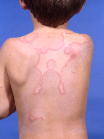
Immune thrombocytopenia
Children usually present with a purpuric rash, but with no systemic upset or organomegaly. Some present with significant coagulopathy.
Platelet transfusions are administered for low platelet counts. Children with significant or life-threatening bleeding, regardless of platelet count, require a combined therapeutic approach with platelet transfusion, corticosteroids, and IVIG.[59]Teachey DT, Lambert MP. Diagnosis and management of autoimmune cytopenias in childhood. Pediatr Clin North Am. 2013 Dec;60(6):1489-511.
http://www.ncbi.nlm.nih.gov/pubmed/24237984?tool=bestpractice.com
Immunoglobulin A (IgA) vasculitis (formerly known as Henoch-Schonlein purpura)
Classic presentation is a tetrad of petechial or purpuric lesions (typically on lower extremities), abdominal pain, arthritis/arthralgia, and IgA nephropathy.[86]Mathur AN, Mathes EF. Urticaria mimickers in children. Dermatol Ther. 2013 Nov-Dec;26(6):467-75.
http://www.ncbi.nlm.nih.gov/pubmed/24552410?tool=bestpractice.com
[87]Tabel Y, Inanc FC, Dogan DG, et al. Clinical features of children with Henoch-Schonlein purpura: risk factors associated with renal involvement. Iran J Kidney Dis. 2012 Jul;6(4):269-74.
http://www.ncbi.nlm.nih.gov/pubmed/22797096?tool=bestpractice.com
In a subpopulation of patients, renal impairment with rapidly progressive nephritis may develop. This generally requires a combination of corticosteroids, immunosuppressants, and plasmapheresis.[86]Mathur AN, Mathes EF. Urticaria mimickers in children. Dermatol Ther. 2013 Nov-Dec;26(6):467-75.
http://www.ncbi.nlm.nih.gov/pubmed/24552410?tool=bestpractice.com
[Figure caption and citation for the preceding image starts]: Palpable purpura on the lower extremities of a child with immunoglobulin A (IgA) vasculitis (formerly known as Henoch-Schonlein purpura)Courtesy of Paul F. Roberts, Mayo Clinic, Jacksonville, FL [Citation ends]. [Figure caption and citation for the preceding image starts]: Immunoglobulin A (IgA) vasculitis (formerly known as Henoch-Schonlein purpura): purpura mainly affects the legs, up to the gluteal muscles, inflaming the joints of the ankle and knee, but can also affect the arms and elbowFrom Wikimedia Commons [Citation ends].
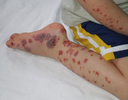
Child abuse and self-harm
The presence of unusual skin marks, multiple bruises of varying ages, recurrent instances of 'unexplained' injury, delayed presentation, and certain types of injuries (metaphyseal 'bucket handle' fractures, long bone fractures, posterior rib fractures) should raise suspicion of child abuse.[88]Kellogg ND; American Academy of Pediatrics Committee on Child Abuse and Neglect. Evaluation of suspected child physical abuse. Pediatrics. 2007 Jun;119(6):1232-41.
https://pediatrics.aappublications.org/content/119/6/1232.long
http://www.ncbi.nlm.nih.gov/pubmed/17545397?tool=bestpractice.com
If child abuse is suspected, child protection procedures should be followed immediately.
Children may cut, pick, or burn their skin often in accessible areas such as the face and limbs. Dermatitis artefacta (DA) is a skin condition caused by the actions of the patient on the skin, hair, nails, or mucosae.[64]Mohandas P, Ravenscroft JC, Bewley A. Dermatitis artefacta in childhood and adolescence: a spectrum of disease. G Ital Dermatol Venereol. 2018 Aug;153(4):525-34.
http://www.ncbi.nlm.nih.gov/pubmed/29683292?tool=bestpractice.com
Deliberate self‐harm differs from DA in that patients will often take responsibility for self-harm but will not acknowledge their actions in DA. This is an indicator of mental health problems and/or social problems and urgent referral to a mental health professional is warranted.
Kawasaki disease
Kawasaki disease is an acute, multi-system, febrile disease that primarily affects children aged <5 years. The rash is usually maculopapular, with diffuse erythroderma, or erythema multiforme-like; perineal erythema and desquamation is particularly pronounced.[89]McCrindle BW, Rowley AH, Newburger JW, et al. Diagnosis, treatment, and long-term management of Kawasaki disease: a scientific statement for health professionals from the American Heart Association. Circulation. 2017 Apr 25;135(17):e927-99.
https://www.ahajournals.org/doi/10.1161/CIR.0000000000000484
http://www.ncbi.nlm.nih.gov/pubmed/28356445?tool=bestpractice.com
[90]Jone PN, Tremoulet A, Choueiter N, et al. Update on diagnosis and management of Kawasaki disease: a scientific statement from the American Heart Association. Circulation. 2024 Dec 3;150(23):e481-500.
https://www.ahajournals.org/doi/full/10.1161/CIR.0000000000001295?rfr_dat=cr_pub++0pubmed&url_ver=Z39.88-2003&rfr_id=ori%3Arid%3Acrossref.org
http://www.ncbi.nlm.nih.gov/pubmed/39534969?tool=bestpractice.com
Diagnostic criteria include high fever for ≥4 days, without any other explanation, plus at least four of the following five principal criteria:[89]McCrindle BW, Rowley AH, Newburger JW, et al. Diagnosis, treatment, and long-term management of Kawasaki disease: a scientific statement for health professionals from the American Heart Association. Circulation. 2017 Apr 25;135(17):e927-99.
https://www.ahajournals.org/doi/10.1161/CIR.0000000000000484
http://www.ncbi.nlm.nih.gov/pubmed/28356445?tool=bestpractice.com
[90]Jone PN, Tremoulet A, Choueiter N, et al. Update on diagnosis and management of Kawasaki disease: a scientific statement from the American Heart Association. Circulation. 2024 Dec 3;150(23):e481-500.
https://www.ahajournals.org/doi/full/10.1161/CIR.0000000000001295?rfr_dat=cr_pub++0pubmed&url_ver=Z39.88-2003&rfr_id=ori%3Arid%3Acrossref.org
http://www.ncbi.nlm.nih.gov/pubmed/39534969?tool=bestpractice.com
Bilateral conjunctival injection without exudate
Cervical lymphadenopathy (usually unilateral)
Oropharyngeal changes (including hyperaemia, oral fissures, strawberry tongue)
Peripheral extremity changes (including erythema, oedema in the acute phase, and desquamation of hands and feet in the subacute phase)
Polymorphous rash (maculopapular, diffuse erythroderma, or erythema multiforme-like)
Experienced clinicians may establish the diagnosis earlier, at 3 days of fever.[90]Jone PN, Tremoulet A, Choueiter N, et al. Update on diagnosis and management of Kawasaki disease: a scientific statement from the American Heart Association. Circulation. 2024 Dec 3;150(23):e481-500.
https://www.ahajournals.org/doi/full/10.1161/CIR.0000000000001295?rfr_dat=cr_pub++0pubmed&url_ver=Z39.88-2003&rfr_id=ori%3Arid%3Acrossref.org
http://www.ncbi.nlm.nih.gov/pubmed/39534969?tool=bestpractice.com
The Single Hub and Access point for paediatric Rheumatology in Europe (SHARE) guideline recommends that a diagnosis can be reached before 5 days of fever if four principal criteria are met or if there is evidence of coronary artery dilation (Z score >2 but <2.5) or aneurysm (Z score ≥2.5), or if there is evidence of persistent inflammation, with no alternative diagnosis, and a clinical suspicion of KD.[91]de Graeff N, Groot N, Ozen S, et al. European consensus-based recommendations for the diagnosis and treatment of Kawasaki disease - the SHARE initiative. Rheumatology (Oxford). 2019 Apr 1;58(4):672-82.
http://www.ncbi.nlm.nih.gov/pubmed/30535127?tool=bestpractice.com
Guidance from the UK National Institute for Health and Care Excellence suggests that a child with fever lasting ≥5 days should be assessed for Kawasaki disease.[92]National Institute for Health and Care Excellence. Fever in under 5s: assessment and initial management. Nov 2021 [internet publication].
https://www.nice.org.uk/guidance/ng143
Recognising Kawasaki disease is critical due to the possibly life-threatening complications of untreated Kawasaki disease (e.g., myocarditis, coronary artery dilation).[90]Jone PN, Tremoulet A, Choueiter N, et al. Update on diagnosis and management of Kawasaki disease: a scientific statement from the American Heart Association. Circulation. 2024 Dec 3;150(23):e481-500.
https://www.ahajournals.org/doi/full/10.1161/CIR.0000000000001295?rfr_dat=cr_pub++0pubmed&url_ver=Z39.88-2003&rfr_id=ori%3Arid%3Acrossref.org
http://www.ncbi.nlm.nih.gov/pubmed/39534969?tool=bestpractice.com
[93]Jamieson N, Singh-Grewal D. Kawasaki disease: a clinician's update. Int J Pediatr. 2013;2013:645391.
https://www.hindawi.com/journals/ijpedi/2013/645391
http://www.ncbi.nlm.nih.gov/pubmed/24282419?tool=bestpractice.com
Urgent hospitalisation to address cardiac issues is required, with prompt treatment with IVIG and high-dose aspirin initially, and further treatment as required.[89]McCrindle BW, Rowley AH, Newburger JW, et al. Diagnosis, treatment, and long-term management of Kawasaki disease: a scientific statement for health professionals from the American Heart Association. Circulation. 2017 Apr 25;135(17):e927-99.
https://www.ahajournals.org/doi/10.1161/CIR.0000000000000484
http://www.ncbi.nlm.nih.gov/pubmed/28356445?tool=bestpractice.com
[90]Jone PN, Tremoulet A, Choueiter N, et al. Update on diagnosis and management of Kawasaki disease: a scientific statement from the American Heart Association. Circulation. 2024 Dec 3;150(23):e481-500.
https://www.ahajournals.org/doi/full/10.1161/CIR.0000000000001295?rfr_dat=cr_pub++0pubmed&url_ver=Z39.88-2003&rfr_id=ori%3Arid%3Acrossref.org
http://www.ncbi.nlm.nih.gov/pubmed/39534969?tool=bestpractice.com
Diagnosis is made clinically, as no specific diagnostic test is available. However, an echocardiogram should be obtained in all patients to assess cardiac status and to assess for the presence of coronary artery abnormality.[89]McCrindle BW, Rowley AH, Newburger JW, et al. Diagnosis, treatment, and long-term management of Kawasaki disease: a scientific statement for health professionals from the American Heart Association. Circulation. 2017 Apr 25;135(17):e927-99.
https://www.ahajournals.org/doi/10.1161/CIR.0000000000000484
http://www.ncbi.nlm.nih.gov/pubmed/28356445?tool=bestpractice.com
[90]Jone PN, Tremoulet A, Choueiter N, et al. Update on diagnosis and management of Kawasaki disease: a scientific statement from the American Heart Association. Circulation. 2024 Dec 3;150(23):e481-500.
https://www.ahajournals.org/doi/full/10.1161/CIR.0000000000001295?rfr_dat=cr_pub++0pubmed&url_ver=Z39.88-2003&rfr_id=ori%3Arid%3Acrossref.org
http://www.ncbi.nlm.nih.gov/pubmed/39534969?tool=bestpractice.com
Severe dengue fever
Severe dengue fever is a potentially deadly complication of dengue virus infection and should be addressed promptly. Patients should be hospitalised. Severe dengue is characterised by plasma leakage, fluid accumulation, respiratory distress, severe bleeding, or organ impairment. Symptoms include abdominal pain, persistent vomiting, tachypnoea, bleeding from gums, and haematemesis. These usually develop 3 to 7 days after the first symptoms of the virus, after the fever decreases.[94]World Health Organization. Dengue and severe dengue. Mar 2023 [internet publication].
https://www.who.int/news-room/fact-sheets/detail/dengue-and-severe-dengue
Patients may have fine petechiae scattered on the extremities, axillae, face, and soft palate, usually seen in the febrile period.[1]Knöpfel N, Noguera-Morel L, Latour I, et al. Viral exanthems in children: a great imitator. Clin Dermatol. 2019 May - Jun;37(3):213-26.
http://www.ncbi.nlm.nih.gov/pubmed/31178104?tool=bestpractice.com
[11]Karnad DR, Richards GA, Silva GS, et al. Tropical diseases in the ICU: a syndromic approach to diagnosis and treatment. J Crit Care. 2018 Aug;46:119-26.
http://www.ncbi.nlm.nih.gov/pubmed/29625787?tool=bestpractice.com
Haemorrhagic signs include petechiae, purpura, or a positive tourniquet test (performed by inflating a blood pressure cuff to a point midway between systolic and diastolic blood pressures for 5 minutes; the test is positive if ≥10 petechiae per square inch appear on the forearm). More significant haemorrhage can manifest as epistaxis, gingival bleeding, haematemesis, melaena, or bleeding from a venipuncture site.[95]World Health Organization, Special Programme for Research and Training in Tropical Diseases (TDR). Dengue: guidelines for diagnosis, treatment, prevention and control. April 2009 [internet publication].
https://www.who.int/publications/i/item/9789241547871
Hepatomegaly may be present. Clinical evidence of plasma leakage includes the presence of ascites, postural dizziness, or pleural effusion. Tachycardia, cold clammy skin, and capillary refill time longer than 3 seconds indicate shock.[95]World Health Organization, Special Programme for Research and Training in Tropical Diseases (TDR). Dengue: guidelines for diagnosis, treatment, prevention and control. April 2009 [internet publication].
https://www.who.int/publications/i/item/9789241547871
An FBC should be ordered initially in all symptomatic patients. Typically, leukopenia and thrombocytopenia occur as early as the second day of fever.[95]World Health Organization, Special Programme for Research and Training in Tropical Diseases (TDR). Dengue: guidelines for diagnosis, treatment, prevention and control. April 2009 [internet publication].
https://www.who.int/publications/i/item/9789241547871
Early diagnosis and optimal clinical management reduce the associated morbidity and mortality. Treatment is supportive, as there is no specific antiviral therapy available for the treatment of dengue infection. The only recognised treatment is fluid replacement therapy.[95]World Health Organization, Special Programme for Research and Training in Tropical Diseases (TDR). Dengue: guidelines for diagnosis, treatment, prevention and control. April 2009 [internet publication].
https://www.who.int/publications/i/item/9789241547871