Images and videos
Images


Assessment of rash in children
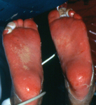
Stevens-Johnson syndrome: epidermal loss on soles of feet
From the personal collection of Dr A. Kowal-Vern
See this image in context in the following section/s:
Assessment of rash in children
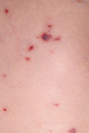
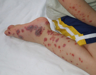
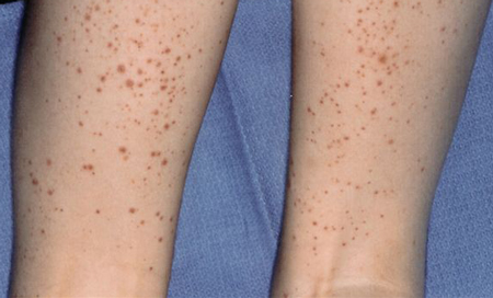
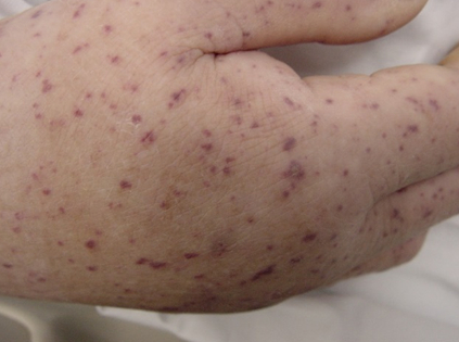
Palpable purpura on the lower extremities of a child with immunoglobulin A (IgA) vasculitis (formerly known as Henoch-Schonlein purpura)
Courtesy of Paul F. Roberts, Mayo Clinic, Jacksonville, FL
See this image in context in the following section/s:

Assessment of rash in children
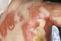
Stevens-Johnson syndrome: targetoid lesions and epidermal loss
From the personal collection of Dr A. Kowal-Vern
See this image in context in the following section/s:

Assessment of rash in children
Toxic epidermal necrolysis with epidermal loss, ocular involvement, and ecthyma gangrenosum
From the personal collection of Dr A. Kowal-Vern
See this image in context in the following section/s:
Assessment of rash in children
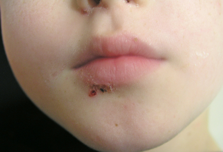
Facial impetigo
Courtesy of Michael Freeman, Bond University, Queensland, Australia
See this image in context in the following section/s:

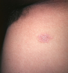
Assessment of rash in children
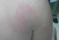
Herald patch
Courtesy of Patricia Treadwell, Riley Hospital for Children, Indianapolis
See this image in context in the following section/s:
Assessment of rash in children
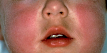
Typical erythematous slapped cheeks of erythema infectiosum
Courtesy of Gary A. Dyer
See this image in context in the following section/s:

Assessment of rash in children
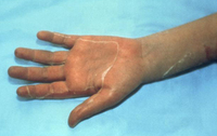
Marked desquamation of the left palm due to toxic shock syndrome, which develops late in the disease
From the CDC and the Public Health Image Library
See this image in context in the following section/s:

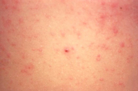
Assessment of rash in children
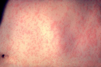
Maculopapular inflammatory rash on the abdomen due to scabies. Of importance, is how this rash appears to resemble many other breakouts, including measles and chickenpox
CDC/Joe Miller
See this image in context in the following section/s:
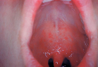
Assessment of rash in children
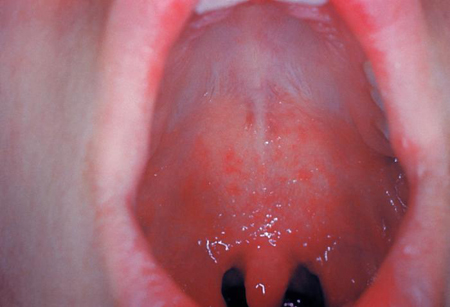
Koplik spots
CDC
See this image in context in the following section/s:
Assessment of rash in children
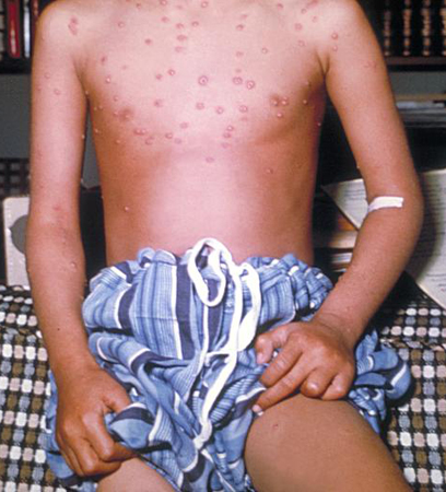
Varicella lesions in young boy
CDC/J.D. Millar and the Public Health Image Library
See this image in context in the following section/s:
Assessment of rash in children
Hypersensitivity rash due to penicillin
CDC Public Health Image Library
See this image in context in the following section/s:

Assessment of rash in children
Petechial rash in invasive meningococcal disease
Thomas AE, et al. BMJ. 2016 Mar 22;352:i1285
See this image in context in the following section/s:

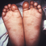
Assessment of rash in children
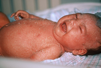
This newborn presented with symptoms of congenital syphilis that included lesions on the soles of both feet
CDC: PHIL image ID 4148
See this image in context in the following section/s:

Assessment of rash in children
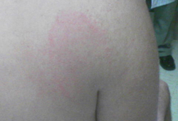
Typical appearance of erythema marginatum on the back of a child with acute rheumatic fever
Courtesy of Professor Mike South, Royal Children’s Hospital Melbourne; used with permission
See this image in context in the following section/s:
Assessment of rash in children
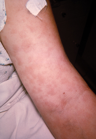
Acute atopic dermatitis in the antecubital fossa of a 9-year-old girl
Courtesy of Christina M. Gelbard, University of Texas, Houston, TX
See this image in context in the following section/s:

Assessment of rash in children
The scarlet fever rash first appears as tiny red bumps on the chest and abdomen that may spread all over the body; looking like sunburn, the skin feels like a rough piece of sandpaper and rash lasts about 2 to 5 days
CDC Public Health Image Library
See this image in context in the following section/s:
Assessment of rash in children
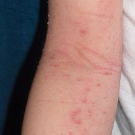
Erythema migrans in Lyme disease
Courtesy of Christian Speil, Southern Illinois University School of Medicine, Springfield, IL
See this image in context in the following section/s:

Assessment of rash in children
Staphylococcal scalded skin syndrome
Dermpics / Science Source / Science Photo Library; reproduced with permission
See this image in context in the following section/s:
Assessment of rash in children
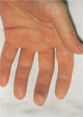
Janeway lesions
From the collection of Sanjay Sharma, St George’s University of London, UK; used with permission
See this image in context in the following section/s:
Assessment of rash in children
Osler node
From the collection of Sanjay Sharma, St George’s University of London, UK; used with permission
See this image in context in the following section/s:

Assessment of rash in children
Child with measles showing the characteristic red blotchy rash on his buttocks and back during the third day of the rash
CDC
See this image in context in the following section/s:

Assessment of rash in children
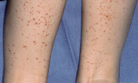
Immunoglobulin A (IgA) vasculitis (formerly known as Henoch-Schonlein purpura): purpura mainly affects the legs, up to the gluteal muscles, inflaming the joints of the ankle and knee, but can also affect the arms and elbow
From Wikimedia Commons
See this image in context in the following section/s:

Assessment of rash in children
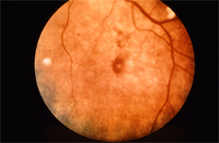
Roth spots
From the collection of Sanjay Sharma, St George’s University of London, UK; used with permission
See this image in context in the following section/s:
Assessment of rash in children
Maculopapular and petechial rash of Rocky Mountain spotted fever
From the collection of Dr Christopher A. Ohl
See this image in context in the following section/s:

Assessment of rash in children
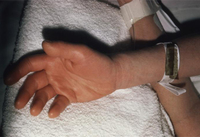
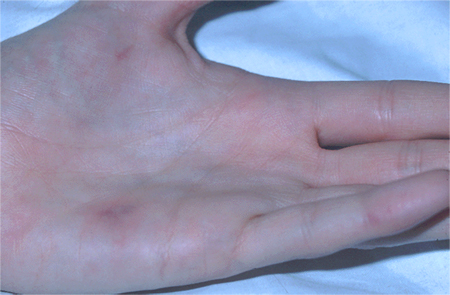
Rash and subcutaneous oedema of the right hand due to toxic shock syndrome
From the CDC and the Public Health Image Library
See this image in context in the following section/s:
Use of this content is subject to our disclaimer