Images and videos
Images

Osteoporosis
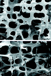
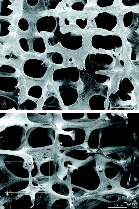
Scanning electron micrographs showing the structure of L3 vertebra in a woman aged 31 years (top) and in a woman aged 70 years (bottom). Note that many of the plate-like structures have become converted to thin rods
Poole KES, et al. BMJ 2006; 333: 1251; used with permission
See this image in context in the following section/s:

Osteoporosis
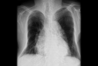
Chest x-ray showing marked deformity and volume loss of bony thorax in a patient with osteoporosis
BMJ Case Reports 2009; doi:10.1136/bcr.07.2008.0359. Copyright ©BMJ publishing group 2010
See this image in context in the following section/s:

Osteoporosis
Management of glucocorticoid-induced osteoporosis
Created by BMJ Knowledge Centre
See this image in context in the following section/s:

Osteoporosis
Management of postmenopausal women with non-glucocorticoid-induced osteoporosis
Created by BMJ Knowledge Centre
See this image in context in the following section/s:
Use of this content is subject to our disclaimer