A thorough clinical assessment is important in diagnosis as the condition is asymptomatic until fracture occurs. Clinical evaluation is based on a history of risk factors and a plan for screening. A history of prior low-impact fragility fracture is an indicator for this condition. Laboratory tests are carried out to exclude reversible secondary causes of low bone mass.
Clinical evaluation
The key components of the patient's history include age, sex, ethnicity, whether women are postmenopausal, history of immobilisation, and whether the patient has a history of previous fractures.[31]ACOG Committee on Clinical Practice Guidelines–Gynecology. Osteoporosis prevention, screening, and diagnosis: ACOG clinical practice guideline no. 1. Obstet Gynecol. 2021 Sep 1;138(3):494-506.
http://www.ncbi.nlm.nih.gov/pubmed/34412075?tool=bestpractice.com
Other risk factors include a family history of maternal hip fracture, secondary amenorrhoea, primary hypogonadism, low calcium intake, vitamin D deficiency, diabetes, rheumatoid arthritis, sarcopenia, and hyperthyroidism.[31]ACOG Committee on Clinical Practice Guidelines–Gynecology. Osteoporosis prevention, screening, and diagnosis: ACOG clinical practice guideline no. 1. Obstet Gynecol. 2021 Sep 1;138(3):494-506.
http://www.ncbi.nlm.nih.gov/pubmed/34412075?tool=bestpractice.com
[41]Chen S, Xu X, Gong H, et al. Global epidemiological features and impact of osteosarcopenia: a comprehensive meta-analysis and systematic review. J Cachexia Sarcopenia Muscle. 2024 Feb;15(1):8-20.
https://pmc.ncbi.nlm.nih.gov/articles/PMC10834350
http://www.ncbi.nlm.nih.gov/pubmed/38086772?tool=bestpractice.com
[45]Lakatos P. Thyroid hormones: beneficial or deleterious for bone? Calcif Tissue Int. 2003 Sep;73(3):205-9.
http://www.ncbi.nlm.nih.gov/pubmed/14667131?tool=bestpractice.com
[67]Qaseem A, Hicks LA, Etxeandia-Ikobaltzeta I, et al. Pharmacologic treatment of primary osteoporosis or low bone mass to prevent fractures in adults: a living clinical guideline from the American College of Physicians. Ann Intern Med. 2023 Feb;176(2):224-38.
https://www.acpjournals.org/doi/full/10.7326/M22-1034
http://www.ncbi.nlm.nih.gov/pubmed/36592456?tool=bestpractice.com
The use of tobacco, alcohol, heparin, anticonvulsants, glucocorticoids, aromatase inhibitors (women) or androgen deprivation treatment (men), proton-pump inhibitors, and drugs associated with an increased risk of falling (e.g., benzodiazepines, antipsychotics, non-benzodiazepine benzodiazepine receptor agonists [also known as z-drugs], antidepressants) should be established.[31]ACOG Committee on Clinical Practice Guidelines–Gynecology. Osteoporosis prevention, screening, and diagnosis: ACOG clinical practice guideline no. 1. Obstet Gynecol. 2021 Sep 1;138(3):494-506.
http://www.ncbi.nlm.nih.gov/pubmed/34412075?tool=bestpractice.com
[51]Seppala LJ, Wermelink AMAT, de Vries M, et al. Fall-risk-increasing drugs: a systematic review and meta-analysis: II. Psychotropics. J Am Med Dir Assoc. 2018 Apr;19(4):371.e11-371.e17.
http://www.ncbi.nlm.nih.gov/pubmed/29402652?tool=bestpractice.com
[67]Qaseem A, Hicks LA, Etxeandia-Ikobaltzeta I, et al. Pharmacologic treatment of primary osteoporosis or low bone mass to prevent fractures in adults: a living clinical guideline from the American College of Physicians. Ann Intern Med. 2023 Feb;176(2):224-38.
https://www.acpjournals.org/doi/full/10.7326/M22-1034
http://www.ncbi.nlm.nih.gov/pubmed/36592456?tool=bestpractice.com
The patient may report changes in height (specifically kyphosis), body weight, diet, and pain.[31]ACOG Committee on Clinical Practice Guidelines–Gynecology. Osteoporosis prevention, screening, and diagnosis: ACOG clinical practice guideline no. 1. Obstet Gynecol. 2021 Sep 1;138(3):494-506.
http://www.ncbi.nlm.nih.gov/pubmed/34412075?tool=bestpractice.com
The risk of fracture should be assessed and history of prior falls investigated.[68]Harvey NC, Odén A, Orwoll E, et al. Falls predict fractures independently of FRAX probability: a meta-analysis of the Osteoporotic Fractures In Men (MrOS) study. J Bone Miner Res. 2018 Mar;33(3):510-6.
https://www.ncbi.nlm.nih.gov/pmc/articles/PMC5842893
http://www.ncbi.nlm.nih.gov/pubmed/29220072?tool=bestpractice.com
Physical examination
The examination may reveal low body mass index or spinal kyphosis due to asymptomatic fracture.
The risk of fall and fracture may be assessed by testing the patient's vision, and assessing balance, gait, and lower-extremity strength.
Imaging
Dual-energy x-ray absorptiometry (DXA)
Measurement of bone mineral density (BMD) using DXA is the gold standard test for diagnosing osteoporosis in patients without an osteoporotic fracture.[3]WHO Scientific Group on the Prevention and Management of Osteoporosis. Prevention and management of osteoporosis: report of a WHO scientific group. (WHO technical report series: 921.) Geneva, Switzerland: WHO; 2003.
https://apps.who.int/iris/handle/10665/42841
[31]ACOG Committee on Clinical Practice Guidelines–Gynecology. Osteoporosis prevention, screening, and diagnosis: ACOG clinical practice guideline no. 1. Obstet Gynecol. 2021 Sep 1;138(3):494-506.
http://www.ncbi.nlm.nih.gov/pubmed/34412075?tool=bestpractice.com
[69]American College of Radiology. ACR appropriateness criteria: osteoporosis and bone mineral density. 2022 [internet publication].
https://acsearch.acr.org/docs/69358/Narrative
[70]ACOG Committee on Clinical Practice Guidelines–Gynecology. Management of postmenopausal osteoporosis: ACOG clinical practice guideline no. 2. Obstet Gynecol. 2022 Apr 1;139(4):698-717.
http://www.ncbi.nlm.nih.gov/pubmed/35594133?tool=bestpractice.com
DXA screening of patients with risk factors for osteoporosis will identify cases prior to fracture. However, there is some debate regarding the appropriate use of BMD analysis. For example, in the context of assessment for prevention of osteoporotic fractures, guidance from the National Institute for Health and Care Excellence states that for women aged 75 years or older who have one or more independent clinical risk factors for fracture or indicators of low BMD, a DXA scan may not be required if the clinician considers it to be clinically inappropriate or unfeasible.[71]National Institute for Health and Care Excellence. Raloxifene and teriparatide for the secondary prevention of osteoporotic fragility fractures in postmenopausal women. Feb 2018 [internet publication].
https://www.nice.org.uk/guidance/ta161
Nonetheless, it is generally agreed that those with fragility fractures (low trauma fracture), oestrogen or testosterone deficiency, height loss, radiographic osteopenia, or rheumatoid arthritis (with or without corticosteroid treatment) and those on oral corticosteroid treatment are eligible for BMD analysis.[72]Adams J, Bishop N. DXA in adults and children. In: Rosen CJ, Compston JE, Lian JB, et al, eds. Primer on the metabolic bone diseases and disorders of mineral metabolism. 7th ed. Washington, DC: American Society for Bone and Mineral Research; 2008:152-8.
Vertebral fracture assessment (VFA)
Moderate or severe vertebral fractures, even when asymptomatic, are strong risk factors for subsequent vertebral and non-vertebral fractures.[24]Gregson CL, Armstrong DJ, Bowden J, et al. UK clinical guideline for the prevention and treatment of osteoporosis. Arch Osteoporos. 2022 Apr 5;17(1):58.
https://www.ncbi.nlm.nih.gov/pmc/articles/PMC8979902
http://www.ncbi.nlm.nih.gov/pubmed/35378630?tool=bestpractice.com
DXA-VFA can be performed during the same session as DXA-BMD analysis to provide images of the lumbar spine. DXA-VFA has moderate sensitivity and high specificity for detecting vertebral fractures compared with spinal radiography, with the advantages of reduced radiation exposure, lower cost, and greater convenience.[62]LeBoff MS, Greenspan SL, Insogna KL, et al. The clinician's guide to prevention and treatment of osteoporosis. Osteoporos Int. 2022 Oct;33(10):2049-102.
https://www.ncbi.nlm.nih.gov/pmc/articles/PMC9546973
http://www.ncbi.nlm.nih.gov/pubmed/35478046?tool=bestpractice.com
[73]Lee JH, Lee YK, Oh SH, et al. A systematic review of diagnostic accuracy of vertebral fracture assessment (VFA) in postmenopausal women and elderly men. Osteoporos Int. 2016 May;27(5):1691-9.
http://www.ncbi.nlm.nih.gov/pubmed/26782682?tool=bestpractice.com
[74]Lems WF, Paccou J, Zhang J, et al. Vertebral fracture: epidemiology, impact and use of DXA vertebral fracture assessment in fracture liaison services. Osteoporos Int. 2021 Mar;32(3):399-411.
https://pmc.ncbi.nlm.nih.gov/articles/PMC7929949
http://www.ncbi.nlm.nih.gov/pubmed/33475820?tool=bestpractice.com
In general, vertebral imaging (with either DXA-VFA or x-ray) should be considered for postmenopausal women and older men with low BMD, height loss, kyphosis, recent or ongoing long-term glucocorticoid treatment, or patients with acute onset back pain and risk factors for osteoporosis.[24]Gregson CL, Armstrong DJ, Bowden J, et al. UK clinical guideline for the prevention and treatment of osteoporosis. Arch Osteoporos. 2022 Apr 5;17(1):58.
https://www.ncbi.nlm.nih.gov/pmc/articles/PMC8979902
http://www.ncbi.nlm.nih.gov/pubmed/35378630?tool=bestpractice.com
[62]LeBoff MS, Greenspan SL, Insogna KL, et al. The clinician's guide to prevention and treatment of osteoporosis. Osteoporos Int. 2022 Oct;33(10):2049-102.
https://www.ncbi.nlm.nih.gov/pmc/articles/PMC9546973
http://www.ncbi.nlm.nih.gov/pubmed/35478046?tool=bestpractice.com
However, recommended indications for vertebral imaging vary; consult local guidance.
Trabecular bone score (TBS)
TBS is a measure of bone microarchitecture derived from lumbar spine DXA images. TBS is an independent predictor of incident fracture and can be used in conjunction with BMD, clinical risk factors, and/or the Fracture Risk Assessment Tool (FRAX) to enhance the accuracy of fracture risk predictions.[69]American College of Radiology. ACR appropriateness criteria: osteoporosis and bone mineral density. 2022 [internet publication].
https://acsearch.acr.org/docs/69358/Narrative
[75]Shevroja E, Reginster JY, Lamy O, et al. Update on the clinical use of trabecular bone score (TBS) in the management of osteoporosis: results of an expert group meeting organized by the European Society for Clinical and Economic Aspects of Osteoporosis, Osteoarthritis and Musculoskeletal Diseases (ESCEO), and the International Osteoporosis Foundation (IOF) under the auspices of WHO Collaborating Center for Epidemiology of Musculoskeletal Health and Aging. Osteoporos Int. 2023 Sep;34(9):1501-29.
https://pmc.ncbi.nlm.nih.gov/articles/PMC10427549
http://www.ncbi.nlm.nih.gov/pubmed/37393412?tool=bestpractice.com
It may be most useful for patients with a BMD T-score or FRAX score close to an intervention threshold.[69]American College of Radiology. ACR appropriateness criteria: osteoporosis and bone mineral density. 2022 [internet publication].
https://acsearch.acr.org/docs/69358/Narrative
[75]Shevroja E, Reginster JY, Lamy O, et al. Update on the clinical use of trabecular bone score (TBS) in the management of osteoporosis: results of an expert group meeting organized by the European Society for Clinical and Economic Aspects of Osteoporosis, Osteoarthritis and Musculoskeletal Diseases (ESCEO), and the International Osteoporosis Foundation (IOF) under the auspices of WHO Collaborating Center for Epidemiology of Musculoskeletal Health and Aging. Osteoporos Int. 2023 Sep;34(9):1501-29.
https://pmc.ncbi.nlm.nih.gov/articles/PMC10427549
http://www.ncbi.nlm.nih.gov/pubmed/37393412?tool=bestpractice.com
Quantitative ultrasound
Quantitative ultrasound (QUS) of the heel can be used as an alternative if DXA is unavailable, but the same diagnostic criteria defined for DXA cannot be applied to this technique.[3]WHO Scientific Group on the Prevention and Management of Osteoporosis. Prevention and management of osteoporosis: report of a WHO scientific group. (WHO technical report series: 921.) Geneva, Switzerland: WHO; 2003.
https://apps.who.int/iris/handle/10665/42841
[76]Fu Y, Li C, Luo W, et al. Fragility fracture discriminative ability of radius quantitative ultrasound: a systematic review and meta-analysis. Osteoporos Int. 2021 Jan;32(1):23-38.
https://www.ncbi.nlm.nih.gov/pmc/articles/PMC7755656
http://www.ncbi.nlm.nih.gov/pubmed/32728897?tool=bestpractice.com
[77]So E, Rushing C, Prissel MA, et al. The role of secondary imaging techniques for assessing bone mineral density in elderly ankle fractures. J Foot Ankle Surg. 2022 Jan-Feb;61(1):149-56.
http://www.ncbi.nlm.nih.gov/pubmed/34312077?tool=bestpractice.com
QUS is, rather than a method for assessing BMD, a method to measure unknown elements of bone strength. QUS may be a good predictor of population-based fracture, but not individual fractures. Additionally, T-score has been tested only for DXA and cannot be applied to QUS.
X-ray
X-ray may reveal osteopenia and/or fractures (e.g., vertebral fractures), but is not diagnostic of condition.[78]Grados F, Fechtenbaum J, Flipon E, et al. Radiographic methods for evaluating osteoporotic vertebral fractures. Joint Bone Spine. 2009 May;76(3):241-7.
http://www.ncbi.nlm.nih.gov/pubmed/19196531?tool=bestpractice.com
It is used to drive the need for DXA assessment when osteopenia is detected coincidentally. It should be considered in patients with pain in the thoraco-lumbar spine, height loss, or thoracic kyphosis. X-ray is also used in therapeutic trials to assess fracture incidence.[78]Grados F, Fechtenbaum J, Flipon E, et al. Radiographic methods for evaluating osteoporotic vertebral fractures. Joint Bone Spine. 2009 May;76(3):241-7.
http://www.ncbi.nlm.nih.gov/pubmed/19196531?tool=bestpractice.com
[Figure caption and citation for the preceding image starts]: Chest x-ray showing marked deformity and volume loss of bony thorax in a patient with osteoporosisBMJ Case Reports 2009; doi:10.1136/bcr.07.2008.0359. Copyright ©BMJ publishing group 2010 [Citation ends].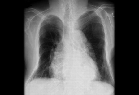
Quantitative CT
Quantitative computed tomography (CT) can be used to measure trabecular bone density if DXA is unavailable.[69]American College of Radiology. ACR appropriateness criteria: osteoporosis and bone mineral density. 2022 [internet publication].
https://acsearch.acr.org/docs/69358/Narrative
[79]Cheng X, Wang L, Wang Q, et al. Validation of quantitative computed tomography-derived areal bone mineral density with dual energy X-ray absorptiometry in an elderly Chinese population. Chin Med J (Engl). 2014;127(8):1445-9.
http://www.ncbi.nlm.nih.gov/pubmed/24762586?tool=bestpractice.com
[80]Cann CE, Adams JE, Brown JK, et al. CTXA hip--an extension of classical DXA measurements using quantitative CT. PLoS One. 2014 Mar 17;9(3):e91904.
https://journals.plos.org/plosone/article?id=10.1371/journal.pone.0091904
http://www.ncbi.nlm.nih.gov/pubmed/24637515?tool=bestpractice.com
Quantitative CT may also be useful for assessing patients with severe degenerative spine disease, obesity, or very tall or short patients.[69]American College of Radiology. ACR appropriateness criteria: osteoporosis and bone mineral density. 2022 [internet publication].
https://acsearch.acr.org/docs/69358/Narrative
Projectional quantitative CT can provide hip BMD measurements equivalent to DXA that may be interpreted using World Health Organization (WHO) T-score criteria, but WHO T-score criteria cannot be applied to spine quantitative CT measurements.[69]American College of Radiology. ACR appropriateness criteria: osteoporosis and bone mineral density. 2022 [internet publication].
https://acsearch.acr.org/docs/69358/Narrative
[81]Royal Australian College of General Practitioners. Osteoporosis management and fracture prevention in post-menopausal women and men >50 years of age. Mar 2024 [internet publication].
https://www.racgp.org.au/clinical-resources/clinical-guidelines/key-racgp-guidelines/view-all-racgp-guidelines/osteoporosis/executive-summary
FRAX
The FRAX was developed by the WHO to assess fracture risk. FRAX integrates clinical risk factors for fracture, with or without BMD scores at the femoral neck, to calculate a 10-year fracture probability for men and women.
FRAX can be used to help assess the need for BMD testing or pharmacological treatment.[24]Gregson CL, Armstrong DJ, Bowden J, et al. UK clinical guideline for the prevention and treatment of osteoporosis. Arch Osteoporos. 2022 Apr 5;17(1):58.
https://www.ncbi.nlm.nih.gov/pmc/articles/PMC8979902
http://www.ncbi.nlm.nih.gov/pubmed/35378630?tool=bestpractice.com
[31]ACOG Committee on Clinical Practice Guidelines–Gynecology. Osteoporosis prevention, screening, and diagnosis: ACOG clinical practice guideline no. 1. Obstet Gynecol. 2021 Sep 1;138(3):494-506.
http://www.ncbi.nlm.nih.gov/pubmed/34412075?tool=bestpractice.com
[81]Royal Australian College of General Practitioners. Osteoporosis management and fracture prevention in post-menopausal women and men >50 years of age. Mar 2024 [internet publication].
https://www.racgp.org.au/clinical-resources/clinical-guidelines/key-racgp-guidelines/view-all-racgp-guidelines/osteoporosis/executive-summary
However, the American College of Obstetricians and Gynecologists highlights that the FRAX tool may be limited due to the inability to input specific amounts, dosage, or duration for alcohol intake, corticosteroid use, smoking, or the number of prior fractures. History of recent falls and spine BMD are not incorporated into the model, both of which increase the risk of osteoporotic fracture, which may lead to an underestimation of risk for these patients.[31]ACOG Committee on Clinical Practice Guidelines–Gynecology. Osteoporosis prevention, screening, and diagnosis: ACOG clinical practice guideline no. 1. Obstet Gynecol. 2021 Sep 1;138(3):494-506.
http://www.ncbi.nlm.nih.gov/pubmed/34412075?tool=bestpractice.com
The FRAXplus calculator addresses some of these limitations with the introduction of additional variables (e.g., recency of fragility fracture, duration of type 2 diabetes mellitus, number of falls in the past year) to provide a more accurate estimate of fracture risk.[82]Fuggle NR, Beaudart C, Bruyère O, et al. Evidence-based guideline for the management of osteoporosis in men. Nat Rev Rheumatol. 2024 Apr;20(4):241-51.
https://www.nature.com/articles/s41584-024-01094-9
http://www.ncbi.nlm.nih.gov/pubmed/38485753?tool=bestpractice.com
The American College of Physicians (ACP) notes that many risk assessment tools are available, but suggests an individualised assessment of baseline fracture risk based on bone density, history of fractures, response to prior osteoporosis treatment, and other risk factors.[67]Qaseem A, Hicks LA, Etxeandia-Ikobaltzeta I, et al. Pharmacologic treatment of primary osteoporosis or low bone mass to prevent fractures in adults: a living clinical guideline from the American College of Physicians. Ann Intern Med. 2023 Feb;176(2):224-38.
https://www.acpjournals.org/doi/full/10.7326/M22-1034
http://www.ncbi.nlm.nih.gov/pubmed/36592456?tool=bestpractice.com
Laboratory evaluation
Biochemical markers of bone resorption, in conjunction with BMD, can be useful predictors of fracture risk and are typically ordered to aid in the decision whether to start treatment.[3]WHO Scientific Group on the Prevention and Management of Osteoporosis. Prevention and management of osteoporosis: report of a WHO scientific group. (WHO technical report series: 921.) Geneva, Switzerland: WHO; 2003.
https://apps.who.int/iris/handle/10665/42841
Bone turnover markers can be used specifically in monitoring response to treatment rather than for the approach to treatment.[83]Allende-Vigo MZ. The use of biochemical markers of bone turnover in osteoporosis. P R Health Sci J. 2007 Jun;26(2):91-5.
http://www.ncbi.nlm.nih.gov/pubmed/17722420?tool=bestpractice.com
[84]Vasikaran S, Eastell R, Bruyère O, et al. Markers of bone turnover for the prediction of fracture risk and monitoring of osteoporosis treatment: a need for international reference standards. Osteoporos Int. 2011 Feb;22(2):391-420.
http://www.ncbi.nlm.nih.gov/pubmed/21184054?tool=bestpractice.com
Laboratory evaluation is performed after diagnosis to ensure that there are no underlying causes of disease for which there is clinical suspicion based on history and physical examination.[2]NIH Consensus Development Panel on Osteoporosis Prevention, Diagnosis, and Therapy. Osteoporosis prevention, diagnosis, and therapy. JAMA. 2001 Feb 14;285(6):785-95.
http://www.ncbi.nlm.nih.gov/pubmed/11176917?tool=bestpractice.com
Initial laboratory tests for all patients assess renal function, as well as calcium, albumin, phosphorus, and vitamin D levels. In men, it is also appropriate to check testosterone levels.
Results that suggest an underlying cause of osteoporosis are:[3]WHO Scientific Group on the Prevention and Management of Osteoporosis. Prevention and management of osteoporosis: report of a WHO scientific group. (WHO technical report series: 921.) Geneva, Switzerland: WHO; 2003.
https://apps.who.int/iris/handle/10665/42841
Vitamin D deficiency (defined as 25-hydroxyvitamin D levels of <20 nanograms/mL)
Testosterone deficiency (in men)
Abnormal thyroid hormone levels (could indicate hyperthyroidism)
High levels of parathyroid hormone (could indicate hyperparathyroidism)
High levels of urinary free cortisol (could indicate Cushing's syndrome)
Abnormal serum/urine protein electrophoresis (could indicate myeloma)
Low serum 25-hydroxyvitamin D levels, low serum and urinary calcium levels, and low serum phosphate levels in conjunction with high levels of serum PTH and alkaline phosphatase (could indicate a vitamin D deficiency with or without osteomalacia).