Images and videos
Images
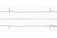


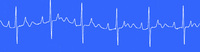

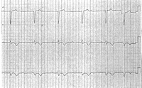

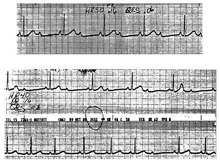
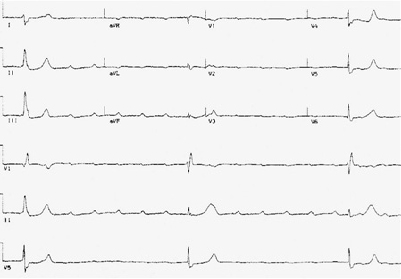
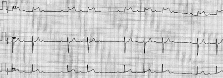
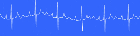
Bradycardia
ECG showing Mobitz II second-degree atrioventricular (AV) block
From the collection of Brian Olshansky, MD, FAHA, FACC, FHRS, FESC; used with permission
See this image in context in the following section/s:
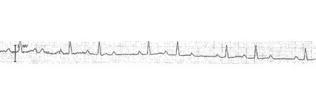
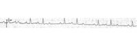
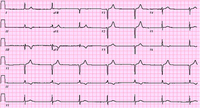
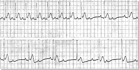
Bradycardia
ECG showing 2:1 atrioventricular (AV) block
From the collection of Brian Olshansky, MD, FAHA, FACC, FHRS, FESC; used with permission
See this image in context in the following section/s:
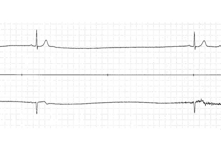
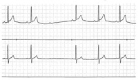
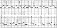
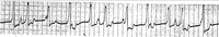
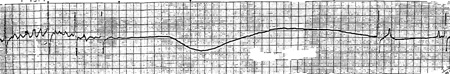
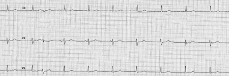
Bradycardia
ECG showing sinus pause
From the collection of Brian Olshansky, MD, FAHA, FACC, FHRS, FESC; used with permission
See this image in context in the following section/s:
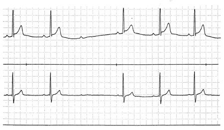
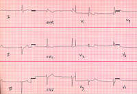
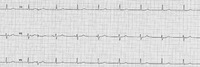
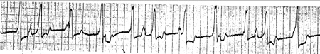
Bradycardia
ECG showing Mobitz II second-degree atrioventricular (AV) block
From the collection of Brian Olshansky, MD, FAHA, FACC, FHRS, FESC; used with permission
See this image in context in the following section/s:
Bradycardia
ECG showing complete atrioventricular (AV) block with junctional escape
From the collection of Brian Olshansky, MD, FAHA, FACC, FHRS, FESC; used with permission
See this image in context in the following section/s:

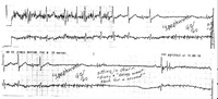
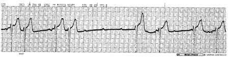
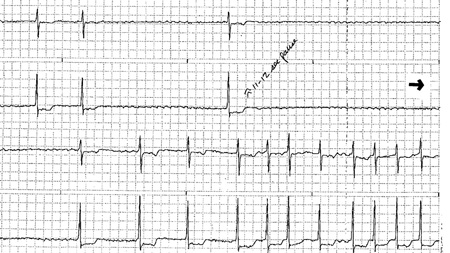
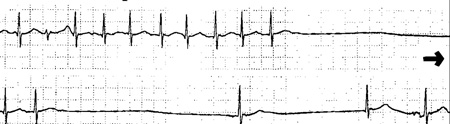
Bradycardia
ECG showing tachy-brady syndrome
From the collection of Brian Olshansky, MD, FAHA, FACC, FHRS, FESC; used with permission
See this image in context in the following section/s:
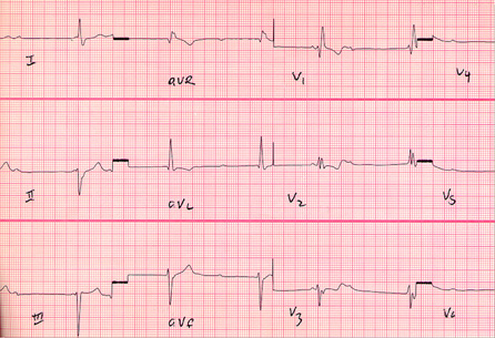
Bradycardia
ECG showing complete atrioventricular (AV) block
From the collection of Brian Olshansky, MD, FAHA, FACC, FHRS, FESC; used with permission
See this image in context in the following section/s:
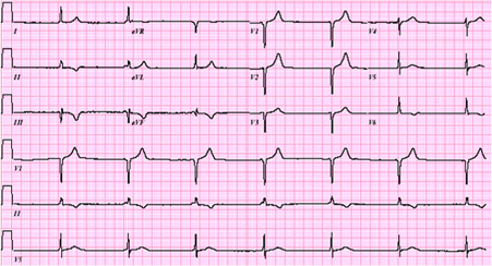
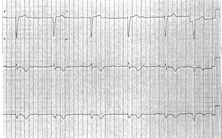
Bradycardia
ECG showing complete atrioventricular (AV) block with ventricular escape
From the collection of Brian Olshansky, MD, FAHA, FACC, FHRS, FESC; used with permission
See this image in context in the following section/s:
Bradycardia
ECG showing junctional rhythm
From the collection of Brian Olshansky, MD, FAHA, FACC, FHRS, FESC; used with permission
See this image in context in the following section/s:
Bradycardia
ECG showing Mobitz I (Wenckebach) second-degree atrioventricular (AV) block during acute inferior wall myocardial infarction
From the collection of Brian Olshansky, MD, FAHA, FACC, FHRS, FESC; used with permission
See this image in context in the following section/s:
Bradycardia
ECG showing tachy-brady syndrome
From the collection of Brian Olshansky, MD, FAHA, FACC, FHRS, FESC; used with permission
See this image in context in the following section/s:
Bradycardia
ECG showing Mobitz I (Wenckebach) second-degree atrioventricular (AV) block
From the collection of Brian Olshansky, MD, FAHA, FACC, FHRS, FESC; used with permission
See this image in context in the following section/s:
Bradycardia
ECG showing complete atrioventricular (AV) block with ventricular escape
From the collection of Brian Olshansky, MD, FAHA, FACC, FHRS, FESC; used with permission
See this image in context in the following section/s:
Bradycardia
ECG showing tachy-brady syndrome
From the collection of Brian Olshansky, MD, FAHA, FACC, FHRS, FESC; used with permission
See this image in context in the following section/s:
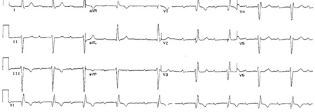
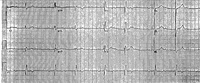
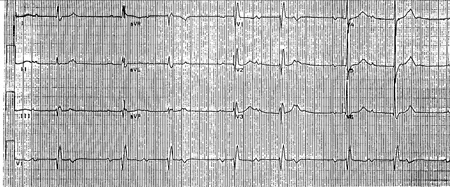
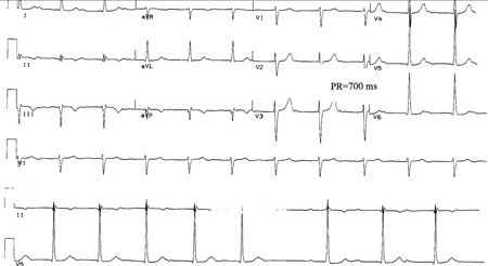
Bradycardia
ECG showing first-degree atrioventricular (AV) block
From the collection of Brian Olshansky, MD, FAHA, FACC, FHRS, FESC; used with permission
See this image in context in the following section/s:
Bradycardia
ECG showing tachy-brady syndrome
From the collection of Brian Olshansky, MD, FAHA, FACC, FHRS, FESC; used with permission
See this image in context in the following section/s:
Bradycardia
ECG showing Mobitz I (Wenckebach) second-degree atrioventricular (AV) block
From the collection of Brian Olshansky, MD, FAHA, FACC, FHRS, FESC; used with permission
See this image in context in the following section/s:
Bradycardia
ECG showing 2:1 atrioventricular (AV) block
From the collection of Brian Olshansky, MD, FAHA, FACC, FHRS, FESC; used with permission
See this image in context in the following section/s:
Bradycardia
ECG showing complete atrioventricular (AV) block
From the collection of Brian Olshansky, MD, FAHA, FACC, FHRS, FESC; used with permission
See this image in context in the following section/s:
Bradycardia
ECG showing interference atrioventricular (AV) dissociation
From the collection of Brian Olshansky, MD, FAHA, FACC, FHRS, FESC; used with permission
See this image in context in the following section/s:
Bradycardia
ECG showing interference atrioventricular (AV) dissociation
From the collection of Brian Olshansky, MD, FAHA, FACC, FHRS, FESC; used with permission
See this image in context in the following section/s:
Videos
 How to perform an ECG animated demonstration
How to record an ECG. Demonstrates placement of chest and limb electrodes.
Use of this content is subject to our disclaimer