Images and videos
Images
Gastro-oesophageal reflux disease
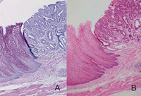
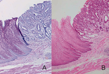
Barrett's segment lined by gastric-type epithelium without intestinal metaplasia in the form of goblet cells (magnification ×100). (A) Mucin staining using Alcian Blue-Periodic Acid Schiff showing neutral magenta colour (mucin of gastric epithelial type); (B) H&E staining reveals a Barrett’s oesophagus segment lined by gastric-type epithelium without intestinal metaplasia but with features of high-grade dysplasia
Lieberman ELB, Lao-Sirieix P, Saeed I, et al. The definition and management of Barrett’s oesophagus: a case report, review of the literature and a suggestion for the future. BMJ Case Reports. 2009; doi:10.1136/bcr.07.2008.0450
See this image in context in the following section/s:
Gastro-oesophageal reflux disease
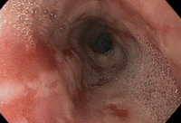
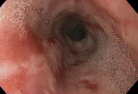
Moderate to severe oesophagitis with multiple linear, clean-based oesophageal ulcers
From the collection of Dr Douglas G. Adler; used with permission
See this image in context in the following section/s:

Gastro-oesophageal reflux disease
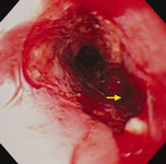
Upper gastrointestinal endoscopy revealing a fistula related to oesophageal carcinoma (arrow)
Wang S-C, Tseng J-C, Lee R-M, et al. Tracheo-oesophageal fistula in a patient with oesophageal squamous cell carcinoma. BMJ Case Reports. 2009; doi:10.1136/bcr.09.2008.0865
See this image in context in the following section/s:
Use of this content is subject to our disclaimer