Investigations
1st investigations to order
electrocardiogram (ECG)
Test
Infants with severe coarctation may show right ventricular hypertrophy (RVH) .
Children or adults with undiagnosed or unrepaired coarctation may show left ventricular hypertrophy (LVH).
A normal ECG should not preclude an echocardiogram or referral to a paediatric cardiologist if the 4 extremity blood pressures or lower extremity pulses are abnormal.
Result
may be normal; show RVH or LVH
chest x-ray (CXR)
Test
Infants with severe coarctation may show cardiomegaly and increased pulmonary vascularity due to pulmonary venous congestion.
Older children and adults with undiagnosed or unrepaired coarctation may show posterior rib notching from collateral vessel enlargement.
A '3' sign may be seen at the site of the coarctation, representing the pre- and post-coarctation dilation.
In a patient with physical findings suspicious for aortic coarctation, a CXR is not necessary before echocardiogram or referral to a paediatric cardiologist.
Result
age and severity dependent; may be normal, have cardiomegaly, or show posterior rib notching
echocardiogram
Test
High parasternal or suprasternal notch view will typically show the discrete narrowing in the thoracic aorta.[Figure caption and citation for the preceding image starts]: Two-dimensional suprasternal notch view of the aortic arch. Focal narrowing of the aortic arch (arrow) in the typical juxtaductal regionFrom the personal collection of Jeffrey Gossett, MD, Children's Memorial Hospital, Northwestern University, Chicago; used with permission [Citation ends].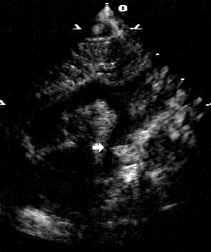 Colour Doppler and pulse Doppler allow calculation of the pressure gradient across the narrowed segment.[Figure caption and citation for the preceding image starts]: Addition of colour Doppler shows turbulent higher velocity flow after the obstructed areaFrom the personal collection of Jeffrey Gossett, MD, Children's Memorial Hospital, Northwestern University, Chicago; used with permission [Citation ends].
Complete two-dimensional and Doppler echocardiogram can accurately establish the diagnosis of aortic coarctation in most patients as well as determining any associated malformations.
In neonates with critical coarctation, the patency of the ductus can be assessed. Additionally, continuous Doppler of the abdominal aorta will often show diastolic run off.[21]
Result
discrete narrowing in the thoracic aorta; pressure gradient across narrowing
Investigations to consider
computed tomography (CT) angiography
Test
CT angiography is a useful adjunct to echocardiography to fully define the anatomy of the aortic arch, especially in older children and adults.[19][22]
Three-dimensional reconstructions can augment the understanding of the anatomy of the narrowing.[23][Figure caption and citation for the preceding image starts]: Three-dimensional computed tomographic reconstruction visualised from posterior to anterior. The area of coarctation is well seen (arrow)From the personal collection of Jeffrey Gossett, MD, Children's Memorial Hospital, Northwestern University, Chicago; used with permission [Citation ends].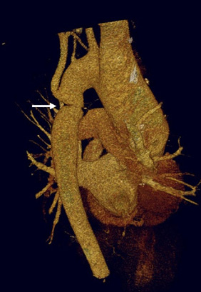
Older patients diagnosed with aortic coarctation should also be screened for intracranial aneurysms by CT angiography.[20]
Result
abnormal anatomy of aortic arch
magnetic resonance imaging (MRI)/magnetic resonance angiography (MRA)
Test
MRI/MRA is another useful adjunctive imaging modality that can help fully delineate the three-dimensional arch anatomy.[19] Advantages include the lack of ionising radiation, but in younger children anaesthesia may be required.[6][22][23]
MRA can be used to screen for intracranial aneurysms in older patients with a history of coarctation of the aorta.[20]
Result
abnormal anatomy of aortic arch
cardiac catheterisation
Test
Cardiac catheterisation is not typically necessary for diagnosing aortic coarctation. While direct measurement of the gradient across the aortic coarctation is obtained with cardiac catheterisation, this is usually performed with the intention to intervene and repair a coarcted segment, either by balloon angioplasty or stent expansion.[19][Figure caption and citation for the preceding image starts]: Angiography in the ascending aorta shows a focal area of narrowing after the left subclavian arteryFrom the personal collection of Jeffrey Gossett, MD, Children's Memorial Hospital, Northwestern University, Chicago; used with permission [Citation ends].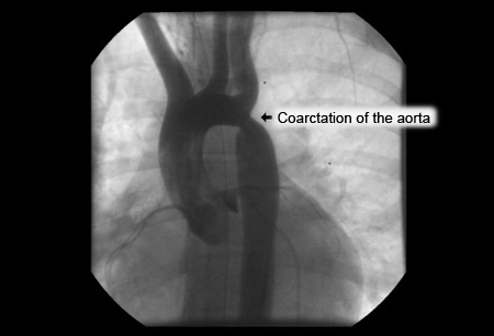 [Figure caption and citation for the preceding image starts]: After stent placement the narrowed area is markedly improvedFrom the personal collection of Jeffrey Gossett, MD, Children's Memorial Hospital, Northwestern University, Chicago; used with permission [Citation ends].
Result
abnormal gradient across narrowing; therapeutic intervention possible
Use of this content is subject to our disclaimer