Images and videos
Images


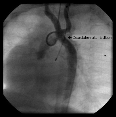
Aortic coarctation
Balloon angioplasty of the narrowed region leads to complete resolution of the obstruction
From the personal collection of Jeffrey Gossett, MD, Children's Memorial Hospital, Northwestern University, Chicago; used with permission
See this image in context in the following section/s:
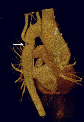
Aortic coarctation
Three-dimensional computed tomographic reconstruction visualised from posterior to anterior. The area of coarctation is well seen (arrow)
From the personal collection of Jeffrey Gossett, MD, Children's Memorial Hospital, Northwestern University, Chicago; used with permission
See this image in context in the following section/s:
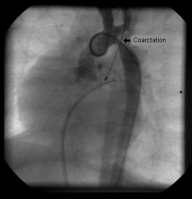
Aortic coarctation
Aortic angiography in an infant after surgical reconstruction of the aortic arch shows marked narrowing at the distal aspect of the surgical repair
From the personal collection of Jeffrey Gossett, MD, Children's Memorial Hospital, Northwestern University, Chicago; used with permission
See this image in context in the following section/s:

Aortic coarctation
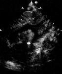
Two-dimensional suprasternal notch view of the aortic arch. Focal narrowing of the aortic arch (arrow) in the typical juxtaductal region
From the personal collection of Jeffrey Gossett, MD, Children's Memorial Hospital, Northwestern University, Chicago; used with permission
See this image in context in the following section/s:

Aortic coarctation
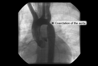
Angiography in the ascending aorta shows a focal area of narrowing after the left subclavian artery
From the personal collection of Jeffrey Gossett, MD, Children's Memorial Hospital, Northwestern University, Chicago; used with permission
See this image in context in the following section/s:

Aortic coarctation
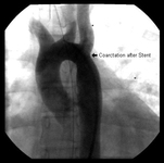
After stent placement the narrowed area is markedly improved
From the personal collection of Jeffrey Gossett, MD, Children's Memorial Hospital, Northwestern University, Chicago; used with permission
See this image in context in the following section/s:

Aortic coarctation
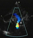
Addition of colour Doppler shows turbulent higher velocity flow after the obstructed area
From the personal collection of Jeffrey Gossett, MD, Children's Memorial Hospital, Northwestern University, Chicago; used with permission
See this image in context in the following section/s:

Aortic coarctation
Abdominal coarctation
N. Pal, D. McEneaney. BMJ Case Reports 2009
See this image in context in the following section/s:
Use of this content is subject to our disclaimer