Approach
Evaluation focuses on historical information about pain location (localised or widespread), precipitants, modifying factors, and comorbid medical illness.
Initial evaluation
Pain location is best identified using a patient-completed pain diagram. [Figure caption and citation for the preceding image starts]: Patients should be asked to complete a pain drawing to determine whether pain is localised or widespread by shading all painful areas, using the following key: ///// = pain; ::::: = numbness; *** = burning or hypersensitivity to touchFrom Marcus DA. Chronic pain. A primary care guide to practical management. Totowa, NJ: Humana Press; 2005 [Citation ends].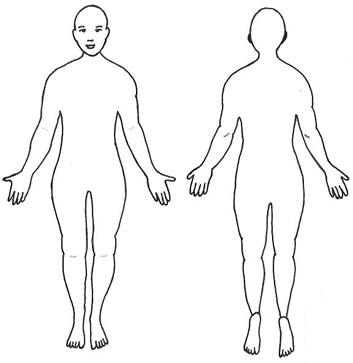
Features of acute pain history that may predispose to developing chronic pain:
Back pain with restricted lumbar flexion
Abnormal neurological examination
Non-localised pain
Insidious onset
Back pain radiating to the lower extremity.
Several factors predispose to the development of chronic pain, and these should be sought in the patient history:[5][6][24][25][26][27]
Female sex
Personal history of trauma or chronic pain; family history of chronic pain
Comorbid mood disorder or psychological distress
Lack of social support
Work dissatisfaction
Nicotine use
Genetic factors (increase predisposition to developing migraine and fibromyalgia)
High-risk occupations (including some healthcare professions such as healthcare assistants, nurses, dentists, and chiropractors; heavy manual workers; car mechanics; housekeepers/caretakers; hairstylists; other occupations that involve prolonged standing; occupations involving sustained low-level movements; soldiers)
Unemployment or previous job change due to pain.
Detailed history
From the initial history it may be possible to determine whether the patient has myofascial, musculoskeletal, or neuropathic pain; fibromyalgia; or a chronic headache syndromes. A more detailed history may then be elicited.
See Diagnostic approach in: Osteoarthritis, Rheumatoid arthritis, Musculoskeletal lower back pain, Complex regional pain syndrome, Diabetic neuropathy, Herpes zoster infection, Fibromyalgia, Migraine headache in adults, Cluster headache, and Tension-type headache.
Myofascial pain
History of muscle tenderness or pain related to physical activity, muscle spasm, and restricted range of motion (ROM) is important.
Disturbances in sleep and mood are common.
Musculoskeletal pain
Chronic back pain is common and may be associated with sensory and motor symptoms, a history of injury or past episodes of acute back pain.
Pain with prolonged walking, relieved by stooping or sitting, may indicate lumbar spinal stenosis due to disc herniation.
Older women with a history of joint and bone pain may have fractures associated with osteoporosis.
The history can help distinguish osteoarthritis (OA) from rheumatoid arthritis (RA).
OA typically affects high-use, weight-bearing, large joints in an asymmetrical fashion. Features of OA include joint pain, morning stiffness in joints, and crepitus with joint movement.
Features of RA include pain and/or swelling in several joints; significant joint stiffness in the morning or after rest; progressive loss of joint function; symmetrical joint involvement; and a good response to non-steroidal anti-inflammatory drugs (NSAIDs).[36]
Polymyalgia rheumatica (female patients older than 50 years of age, with neck, shoulder, and hip pain; rapid response to prednisolone) and polymyositis (symmetrical weakness of shoulder and pelvic girdles) are important musculoskeletal conditions that should be distinguished from fibromyalgia.
Neuropathic pain
Diagnosis of neuropathic pain depends on clinical symptoms and distribution of pain. Hallmarks include burning sensation, hyperalgesia, and allodynia.
Patients may also report areas of numbness or sensory loss in dermatomal distributions or in a stocking/glove pattern. There may be a history of diabetes mellitus or of shingles.
Complex regional pain syndrome often exhibits excessive guarding of the painful extremity and is diagnosed using the International Association for the Study of Pain diagnostic criteria.[37]
Fibromyalgia
Diagnosed using criteria from the American College of Rheumatology:[38]
Widespread pain index (WPI) ≥7 and symptom severity (SS) scale score ≥5, or WPI 3 to 6 and SS scale score ≥9
Symptoms have been present at a similar level for at least 3 months
The patient does not have a disorder that would otherwise explain the pain.
In addition to widespread pain, fibromyalgia patients may have somatic symptoms including chronic headaches, sleep disturbance, depression and/or anxiety, irritable bowel syndrome, genitourinary disturbances, and/or diffuse sensory disturbances.
Chronic headaches
Benign chronically recurring headaches are diagnosed by reviewing headache characteristics. The most important factors in the history are frequency of headaches; duration of attacks; severity of attacks; whether the headache is constant or intermittent, and unilateral or bilateral; associations (nausea, sensitivity to light or noise); and presence of trigger factors or associations with injury or medication overuse.
[Figure caption and citation for the preceding image starts]: Headache diagnostic algorithmMarcus DA. Chronic pain. A primary care guide to practical management. Totowa, NJ: Humana Press; 2005 [Citation ends].
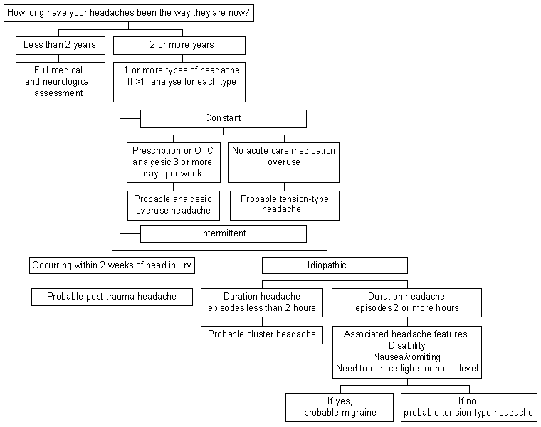
Type of headache is identified using International Classification of Headache Disorders (ICHD)-3 criteria.[2]
Warning symptoms of chronic headache suggesting need for additional work-up:
New headache or significant change in headache character with 2 years
Posterior head or neck pain (may signify trauma or vertebral artery dissection)
Patient more than 50 years of age
Abnormal neurological symptoms.
Examination
Physical capacity evaluation clarifies specific facets of chronic pain disability, is useful for preparing rehabilitation programmes and is particularly beneficial in patients struggling to successfully return to work.
Patients with localised pain are evaluated with physical examination of the muscles and soft tissues (myofascial pain), joints (musculoskeletal pain), and nerves (neuropathic pain), to determine the need for supplemental testing to identify the diagnostic category.
Widespread pain may occur in conjunction with a variety of systemic illnesses. Patients without associated systemic illness should be evaluated to differentiate widespread myofascial pain from fibromyalgia.
Myofascial pain
Diagnosis is based on physical examination and is characterised by restricted active ROM, normal passive ROM, and localised, shortened, and tender muscles with taut bands and trigger points.
Taut band:
Palpable contracted cord-like group of muscle fibres
Point tenderness over taut band
Local twitch response: involuntary contraction of taut band after it is physically plucked or a needle is inserted into it.
Trigger points:
Active if palpation results in pain referred to chronic pain area
Latent if locally tender but without referred pain.
Musculoskeletal pain
Characterised by restricted passive ROM, joint crepitus, joint inflammation, and joint instability. Physical examination features in musculoskeletal pain due to RA.
Joint inflammation (swelling, warmth, tenderness)
Restricted joint ROM
Typically affects small joints in a symmetrical fashion.
Neuropathic pain
Numbness; weakness; reflex changes in distributions consistent with spine, spinal root, or nerve abnormality; abnormal straight leg raising. Allodynia, hyperalgesia, and loss of cold sensation.
Fibromyalgia
Tenderness to palpation over pre-specified points that may not be within areas of pain complaints; full passive ROM and active ROM.
Chronic headache
Further examination is required for patients with concerning headache symptoms:[39]
Complete review of systems
Cervical spine examination
Resting posture
Active ROM
Palpation
Neurological evaluation
Gait
Fundoscopy for papillo-oedema
Assessment of symmetry of face and eye movements
Strength and reflex testing
Sensation to touch.
Imaging
Patients with restricted ROM determined to be related to musculoskeletal/mechanical pain should be evaluated with plain x-rays, which can help distinguish OA (bony spurs, osteophytes and cartilage erosion) from RA (reduced cartilage and inflammatory changes), detect fractures and tumours, and show changes relating to ankylosing spondylitis, fractures associated with osteoporosis, and changes of osteomalacia.
Magnetic resonance imaging (MRI) is sometimes needed for patients with suspected radiculopathy or plexopathy, or for patients with new-onset or concerning headaches to rule out secondary causes (e.g., tumour, ischaemic lesions).
Other investigations
Other investigations are usually not needed but may be necessary in patients with neuropathic pain to determine underlying medical illnesses that may be contributing to peripheral neuropathy.
Electromyogram and nerve conduction velocity studies: may be helpful in identifying mononeuropathies, such as radiculopathy and compressive neuropathies (e.g., carpal tunnel syndrome). Often, abnormalities are also seen in peripheral neuropathy.
Quantitative sensory testing and quantitative sudomotor axon reflex test: frequently used research tools with low diagnostic sensitivity and specificity.[40] Therefore, these tools are not recommended as part of a typical clinical evaluation.
Use of this content is subject to our disclaimer