The most common symptoms of OA are joint pain, stiffness, and sometimes swelling. OA most commonly affects the knee, hip, small hand joints (proximal interphalangeal [PIP] and distal interphalangeal [DIP] joints), and the spine (especially lumbar and cervical regions). In other joints (e.g., the ankle and wrist), OA is rare and there is usually an underlying aetiology (e.g., crystal arthropathy, trauma).
History
More patients present in their 50s as OA is more common at this age, with a higher number of women presenting than men.[9]Zhang Y, Jordan JM. Epidemiology of osteoarthritis. Clin Geriatr Med. 2010 Aug;26(3):355-69.
https://www.ncbi.nlm.nih.gov/pmc/articles/PMC2920533
http://www.ncbi.nlm.nih.gov/pubmed/20699159?tool=bestpractice.com
[10]Prieto-Alhambra D, Judge A, Javaid MK, et al. Incidence and risk factors for clinically diagnosed knee, hip and hand osteoarthritis: influences of age, gender and osteoarthritis affecting other joints. Ann Rheum Dis. 2014 Sep;73(9):1659-64.
https://www.ncbi.nlm.nih.gov/pmc/articles/pmid/23744977
http://www.ncbi.nlm.nih.gov/pubmed/23744977?tool=bestpractice.com
[15]Felson DT, Naimark A, Anderson J, et al. The prevalence of knee osteoarthritis in the elderly: the Framingham Osteoarthritis Study. Arthritis Rheum. 1987;30:914-918.
http://www.ncbi.nlm.nih.gov/pubmed/3632732?tool=bestpractice.com
[16]Felson DT, Zhang Y, Hannan MT, et al. The incidence and natural history of knee osteoarthritis in the elderly. The Framingham Osteoarthritis Study. Arthritis Rheum. 1995;38:1500-1505.
http://www.ncbi.nlm.nih.gov/pubmed/7575700?tool=bestpractice.com
[18]Nevitt MC, Xu L, Zhang Y, et al. Very low prevalence of hip osteoarthritis among Chinese elderly in Beijing, China, compared with whites in the United States: the Beijing osteoarthritis study. Arthritis Rheum. 2002;46:1773-1779.
http://www.ncbi.nlm.nih.gov/pubmed/12124860?tool=bestpractice.com
[19]Zhang Y, Xu L, Nevitt MC, et al. Comparison of the prevalence of knee osteoarthritis between the elderly Chinese population in Beijing and whites in the United States: the Beijing Osteoarthritis Study. Arthritis Rheum. 2001;44:2065-2071.
http://www.ncbi.nlm.nih.gov/pubmed/11592368?tool=bestpractice.com
[24]Spector TD, Cicuttini F, Baker J, et al. Genetic influences on osteoarthritis in women: a twin study. BMJ. 1996;312:940-3.
http://www.ncbi.nlm.nih.gov/pubmed/8616305?tool=bestpractice.com
[56]Zhang W, Doherty M, Leeb BF, et al. EULAR evidence-based recommendations for the diagnosis of hand osteoarthritis: report of a task force of ESCISIT. Ann Rheum Dis. 2009 Jan;68(1):8-17.
https://ard.bmj.com/content/68/1/8.long
http://www.ncbi.nlm.nih.gov/pubmed/18250111?tool=bestpractice.com
Patient history may include a physically demanding job or sport, with joint pain worsening during activities or weight bearing. Joint pain should not be present at night, except in advanced OA; if the patient has joint pain during the night, a differential diagnosis should be considered.
The distribution of joint involvement is important. Some women have inflammatory OA mainly affecting the proximal interphalangeal (PIP) and distal interphalangeal (DIP) joints of the hands, which may be erythematous and swollen. The MCP joint can occasionally be affected by OA; however, if MCP symptoms are present, a differential diagnosis of calcium pyrophosphate dihydrate deposition (CPPD) disease, rheumatoid arthritis, or other secondary aetiology should be considered.[72]Kouki I, Tuffet S, Crema MD, et al. Metacarpophalangeal joint impairment in hand osteoarthritis and its association with mechanical factors: results from the digital cohort osteoarthritis design hand osteoarthritis cohort. Arthritis Care Res (Hoboken). 2022 Oct;74(10):1696-703.
http://www.ncbi.nlm.nih.gov/pubmed/33973396?tool=bestpractice.com
People with OA present with morning stiffness of no more than 30 minutes.[3]Altman R, Asch E, Bloch D, et al; Diagnostic and Therapeutic Criteria Committee of the American Rheumatism Association. Development of criteria for the classification and reporting of osteoarthritis: classification of osteoarthritis of the knee. Arthritis Rheum. 1986;29:1039-49.
http://www.ncbi.nlm.nih.gov/pubmed/3741515?tool=bestpractice.com
[73]National Institute for Health and Care Excellence. Osteoarthritis in over 16s: diagnosis and management. Oct 2022 [internet publication].
https://www.nice.org.uk/guidance/ng226
If stiffness persists for longer than this, other diagnoses should be considered, such as rheumatoid arthritis.
Joint swelling and functional difficulties, such as a knee giving way or locking may be reported by patients. This can reflect an internal derangement, such as a partial meniscal tear or a loose body within the joint.
Physical examination
Weight and body mass index are important, as knee OA and, to a lesser degree, hip OA are common in overweight patients.[23]Reyes C, Leyland KM, Peat G, et al. Association between overweight and obesity and risk of clinically diagnosed knee, hip, and hand osteoarthritis: a population-based cohort study. Arthritis Rheumatol. 2016 Aug;68(8):1869-75.
https://onlinelibrary.wiley.com/doi/10.1002/art.39707
http://www.ncbi.nlm.nih.gov/pubmed/27059260?tool=bestpractice.com
[22]Silverwood V, Blagojevic-Bucknall M, Jinks C, et al. Current evidence on risk factors for knee osteoarthritis in older adults: a systematic review and meta-analysis. Osteoarthritis Cartilage. 2015 Apr;23(4):507-15.
https://www.oarsijournal.com/article/S1063-4584(14)01342-9/fulltext
http://www.ncbi.nlm.nih.gov/pubmed/25447976?tool=bestpractice.com
Swelling may be observed, with bony deformities and malalignment of the affected joint.[33]Felson DT. Clinical practice. Osteoarthritis of the knee. N Engl J Med. 2006;354:841-8.
http://www.ncbi.nlm.nih.gov/pubmed/16495396?tool=bestpractice.com
[48]Sharma L, Song J, Felson DT, et al. The role of knee alignment in disease progression and functional decline in knee osteoarthritis. JAMA. 2001;286:188-195.
http://www.ncbi.nlm.nih.gov/pubmed/11448282?tool=bestpractice.com
[74]van Tunen JAC, Dell'Isola A, Juhl C, et al. Association of malalignment, muscular dysfunction, proprioception, laxity and abnormal joint loading with tibiofemoral knee osteoarthritis - a systematic review and meta-analysis. BMC Musculoskelet Disord. 2018 Jul 28;19(1):273.
https://bmcmusculoskeletdisord.biomedcentral.com/articles/10.1186/s12891-018-2202-8
http://www.ncbi.nlm.nih.gov/pubmed/30055600?tool=bestpractice.com
[32]Chang A, Hayes K, Dunlop D, et al. Thrust during ambulation and progression of knee osteoarthritis. Arthritis Rheum. 2004;50:3897-903.
http://www.ncbi.nlm.nih.gov/pubmed/15593195?tool=bestpractice.com
Bony deformities are particularly common in the hands and lead to enlargement of the PIP joints (Bouchard nodes) and DIP joints (Heberden nodes), as well as squaring at the base of the thumb (the first carpometacarpal joint).[75]Altman R, Alarcon G, Appelrouth D. The American College of Rheumatology criteria for the classification and reporting of osteoarthritis of the hand. Arthritis Rheum. 1990;33:1601-1610.
http://www.ncbi.nlm.nih.gov/pubmed/2242058?tool=bestpractice.com
Bony malalignment is common, particularly in the knee, where OA causes both genu valgum (knock-knees) and genu varum (bow-legs).[33]Felson DT. Clinical practice. Osteoarthritis of the knee. N Engl J Med. 2006;354:841-8.
http://www.ncbi.nlm.nih.gov/pubmed/16495396?tool=bestpractice.com
In addition, a varus thrust, which is a worsening varus alignment in a weight-bearing knee, seems to further worsen the risk of progression of medial knee OA.[32]Chang A, Hayes K, Dunlop D, et al. Thrust during ambulation and progression of knee osteoarthritis. Arthritis Rheum. 2004;50:3897-903.
http://www.ncbi.nlm.nih.gov/pubmed/15593195?tool=bestpractice.com
In advanced knee OA, there may be new bone formation, causing bony swellings around the knee joint.[3]Altman R, Asch E, Bloch D, et al; Diagnostic and Therapeutic Criteria Committee of the American Rheumatism Association. Development of criteria for the classification and reporting of osteoarthritis: classification of osteoarthritis of the knee. Arthritis Rheum. 1986;29:1039-49.
http://www.ncbi.nlm.nih.gov/pubmed/3741515?tool=bestpractice.com
[76]Peat G, Duncan RC, Wood LR, et al. Clinical features of symptomatic patellofemoral joint osteoarthritis. Arthritis Res Ther. 2012 Mar 14;14(2):R63.
https://arthritis-research.biomedcentral.com/articles/10.1186/ar3779
http://www.ncbi.nlm.nih.gov/pubmed/22417687?tool=bestpractice.com
It is common to palpate crepitus during the range of motion of the joint. Limited range of motion, small effusions, and joint line tenderness may be elicited. An abnormal gait can be observed.
Hip examinationA consultant physician demonstrates hip examination techniques, including the Trendelenburg test, the modified Thomas test, and Ober's test. These tests inspect the lateral alignment, anterior and posterior alignment, gait, active and passive movement, and internal and external rotation of the hip.
Knee examinationA consultant physician demonstrates knee examination techniques, inspecting the biomechanics of the knee and assessing for effusion, wasting, or hypertension. The physician performs tests for the integrity of the medial and lateral collateral ligaments, damage to the cruciate ligaments (Lackman's test, Drawer test, Pivot shift test), tests of the patella (patella apprehension test, Clarke's test), and tests of the iliotibial band (Ober's test).
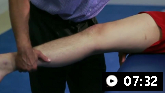
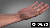
Wrist examinationA consultant physician demonstrates wrist examination techniques for assessing range of movement, inspecting for synovitis, wasting, squaring at the base of the thumb, and previous surgery for carpal or Guyon’s canal syndrome.
OA is essentially a clinical diagnosis
Guidelines recommend that OA is diagnosed clinically based on symptoms, patient age, and examination findings.[3]Altman R, Asch E, Bloch D, et al; Diagnostic and Therapeutic Criteria Committee of the American Rheumatism Association. Development of criteria for the classification and reporting of osteoarthritis: classification of osteoarthritis of the knee. Arthritis Rheum. 1986;29:1039-49.
http://www.ncbi.nlm.nih.gov/pubmed/3741515?tool=bestpractice.com
[73]National Institute for Health and Care Excellence. Osteoarthritis in over 16s: diagnosis and management. Oct 2022 [internet publication].
https://www.nice.org.uk/guidance/ng226
[77]Sakellariou G, Conaghan PG, Zhang W, et al. EULAR recommendations for the use of imaging in the clinical management of peripheral joint osteoarthritis. Ann Rheum Dis. 2017 Sep;76(9):1484-94.
https://ard.bmj.com/content/76/9/1484.long
http://www.ncbi.nlm.nih.gov/pubmed/28389554?tool=bestpractice.com
A clinical diagnosis can be considered in a patient with a typical OA presentation:[3]Altman R, Asch E, Bloch D, et al; Diagnostic and Therapeutic Criteria Committee of the American Rheumatism Association. Development of criteria for the classification and reporting of osteoarthritis: classification of osteoarthritis of the knee. Arthritis Rheum. 1986;29:1039-49.
http://www.ncbi.nlm.nih.gov/pubmed/3741515?tool=bestpractice.com
[73]National Institute for Health and Care Excellence. Osteoarthritis in over 16s: diagnosis and management. Oct 2022 [internet publication].
https://www.nice.org.uk/guidance/ng226
[77]Sakellariou G, Conaghan PG, Zhang W, et al. EULAR recommendations for the use of imaging in the clinical management of peripheral joint osteoarthritis. Ann Rheum Dis. 2017 Sep;76(9):1484-94.
https://ard.bmj.com/content/76/9/1484.long
http://www.ncbi.nlm.nih.gov/pubmed/28389554?tool=bestpractice.com
Atypical features that suggest an alternative or additional diagnosis include:[73]National Institute for Health and Care Excellence. Osteoarthritis in over 16s: diagnosis and management. Oct 2022 [internet publication].
https://www.nice.org.uk/guidance/ng226
Prolonged morning joint-related stiffness
History of recent trauma
Palpable warmth over the joint
Rapid worsening of symptoms
Concerns that may suggest infection or malignancy.
Laboratory assessment
When an alternative or additional diagnosis of rheumatoid arthritis is suspected, inflammatory markers (C-reactive protein [CRP], erythrocyte sedimentation rate [ESR]), rheumatoid factor, and anti-cyclic citrullinated peptide (anti-CCP) antibodies may be helpful as differential tests.[3]Altman R, Asch E, Bloch D, et al; Diagnostic and Therapeutic Criteria Committee of the American Rheumatism Association. Development of criteria for the classification and reporting of osteoarthritis: classification of osteoarthritis of the knee. Arthritis Rheum. 1986;29:1039-49.
http://www.ncbi.nlm.nih.gov/pubmed/3741515?tool=bestpractice.com
[78]Altman R, Alarcon G, Appelrouth D. The American College of Rheumatology criteria for the classification and reporting of osteoarthritis of the hip. Arthritis Rheum. 1991;34:505-514.
http://www.ncbi.nlm.nih.gov/pubmed/2025304?tool=bestpractice.com
These tests are normal in OA.
It should be noted that elevated inflammatory markers are also associated with age, increased weight, and other conditions; therefore, they should not be interpreted as definitive evidence of inflammatory conditions in the absence of other symptoms.
Imaging studies
Imaging studies are not routinely recommended for the diagnosis of OA, they should only be considered if the diagnosis is unclear or an alternative/additional diagnosis is suspected subsequent to initial investigations.[73]National Institute for Health and Care Excellence. Osteoarthritis in over 16s: diagnosis and management. Oct 2022 [internet publication].
https://www.nice.org.uk/guidance/ng226
Radiography
If imaging studies are required, radiography should be considered before other imaging modalities.[77]Sakellariou G, Conaghan PG, Zhang W, et al. EULAR recommendations for the use of imaging in the clinical management of peripheral joint osteoarthritis. Ann Rheum Dis. 2017 Sep;76(9):1484-94.
https://ard.bmj.com/content/76/9/1484.long
http://www.ncbi.nlm.nih.gov/pubmed/28389554?tool=bestpractice.com
[79]Fox MG, Chang EY, Amini B, et al; Expert Panel on Musculoskeletal Imaging. ACR appropriateness criteria®: chronic knee pain. J Am Coll Radiol. 2018 Nov;15(11s):S302-12.
https://www.jacr.org/article/S1546-1440(18)31158-X/fulltext
http://www.ncbi.nlm.nih.gov/pubmed/30392599?tool=bestpractice.com
Conventional radiographic diagnosis of OA includes narrowing of the joint space, osteophytes, subchondral cysts, and subarticular sclerosis.[79]Fox MG, Chang EY, Amini B, et al; Expert Panel on Musculoskeletal Imaging. ACR appropriateness criteria®: chronic knee pain. J Am Coll Radiol. 2018 Nov;15(11s):S302-12.
https://www.jacr.org/article/S1546-1440(18)31158-X/fulltext
http://www.ncbi.nlm.nih.gov/pubmed/30392599?tool=bestpractice.com
[80]Hayashi D, Roemer FW, Guermazi A. Imaging for osteoarthritis. Ann Phys Rehabil Med. 2016 Jun;59(3):161-9.
https://www.sciencedirect.com/science/article/pii/S1877065715005849?via%3Dihub
http://www.ncbi.nlm.nih.gov/pubmed/26797169?tool=bestpractice.com
Radiographs may also help to exclude less common aetiologies for pain and in monitoring unexpected rapid progression or changes in symptoms that may be related to OA severity or an additional diagnosis.[77]Sakellariou G, Conaghan PG, Zhang W, et al. EULAR recommendations for the use of imaging in the clinical management of peripheral joint osteoarthritis. Ann Rheum Dis. 2017 Sep;76(9):1484-94.
https://ard.bmj.com/content/76/9/1484.long
http://www.ncbi.nlm.nih.gov/pubmed/28389554?tool=bestpractice.com
While radiographic evidence of OA is poorly correlated with the symptoms, one study suggested that radiological progression in early symptomatic knee OA over 5 years was related to worsening pain and function.[81]Wesseling J, Bierma-Zeinstra SM, Kloppenburg M, et al. Worsening of pain and function over 5 years in individuals with 'early' OA is related to structural damage: data from the Osteoarthritis Initiative and CHECK (Cohort Hip & Cohort Knee) study. Ann Rheum Dis. 2015;74:347-353.
http://www.ncbi.nlm.nih.gov/pubmed/24243926?tool=bestpractice.com
Magnetic resonance imaging (MRI)
Although more sensitive than plain radiographs in detecting OA changes, MRI is not indicated for the diagnosis of simple OA. If needed, MRI should be ordered if spinal OA with neurological deficits is suspected, to identify and evaluate the extent and severity of spinal stenosis or nerve root entrapment.
MRI can be used to rule out other aetiologies for hip or knee pain, such as avascular necrosis or other less common conditions such as pigmented villonodular synovitis or bone tumours.[77]Sakellariou G, Conaghan PG, Zhang W, et al. EULAR recommendations for the use of imaging in the clinical management of peripheral joint osteoarthritis. Ann Rheum Dis. 2017 Sep;76(9):1484-94.
https://ard.bmj.com/content/76/9/1484.long
http://www.ncbi.nlm.nih.gov/pubmed/28389554?tool=bestpractice.com
[79]Fox MG, Chang EY, Amini B, et al; Expert Panel on Musculoskeletal Imaging. ACR appropriateness criteria®: chronic knee pain. J Am Coll Radiol. 2018 Nov;15(11s):S302-12.
https://www.jacr.org/article/S1546-1440(18)31158-X/fulltext
http://www.ncbi.nlm.nih.gov/pubmed/30392599?tool=bestpractice.com
Although more sensitive than plain x-ray in detecting OA changes, MRI is not indicated for the diagnosis of simple OA in most joints.
Bone marrow lesions (oedema), which are associated with knee pain in OA, are readily detected using MRI.[79]Fox MG, Chang EY, Amini B, et al; Expert Panel on Musculoskeletal Imaging. ACR appropriateness criteria®: chronic knee pain. J Am Coll Radiol. 2018 Nov;15(11s):S302-12.
https://www.jacr.org/article/S1546-1440(18)31158-X/fulltext
http://www.ncbi.nlm.nih.gov/pubmed/30392599?tool=bestpractice.com
[82]Yusuf E, Kortekaas MC, Watt I, et al. Do knee abnormalities visualised on MRI explain knee pain in knee osteoarthritis? A systematic review. Ann Rheum Dis. 2011 Jan;70(1):60-7.
http://www.ncbi.nlm.nih.gov/pubmed/20829200?tool=bestpractice.com
[83]Foong YC, Khan HI, Blizzard L, et al. The clinical significance, natural history and predictors of bone marrow lesion change over eight years. Arthritis Res Ther. 2014 Jul 14;16(4):R149.
https://arthritis-research.biomedcentral.com/articles/10.1186/ar4611
http://www.ncbi.nlm.nih.gov/pubmed/25022807?tool=bestpractice.com
Medial and lateral compartment bone marrow lesions confer increased risk of progression of medial and lateral tibiofemoral OA, respectively.[84]Felson DT, McLaughlin S, Goggins J, et al. Bone marrow edema and its relation to progression to knee osteoarthritis. Ann Intern Med. 2003;139:330-336.
http://www.ncbi.nlm.nih.gov/pubmed/12965941?tool=bestpractice.com
Other imaging modalities
Ultrasound (US) and computed tomography (CT) scans can be used to make additional diagnoses. Soft tissues are best imaged by US or MRI and bone by CT or MRI.[77]Sakellariou G, Conaghan PG, Zhang W, et al. EULAR recommendations for the use of imaging in the clinical management of peripheral joint osteoarthritis. Ann Rheum Dis. 2017 Sep;76(9):1484-94.
https://ard.bmj.com/content/76/9/1484.long
http://www.ncbi.nlm.nih.gov/pubmed/28389554?tool=bestpractice.com
In practice, CT is not widely used for the diagnosis of OA.