Aetiology
The aetiology of most cases of intussusception is unclear but may be related to hyperplasia of Peyer's patches and lymphoid tissue in the intestinal wall resulting from antecedent viral infection. These enlarged lymph nodes may act as the lead point in idiopathic intussusception.[1] Symptoms of an antecedent viral illness are identified in approximately 25% of cases of intussusception.[3][11]
Intussusception in older children and adults is rare and is almost always caused by a pathological lead point. Pathological lead points are anatomical abnormalities of the intestine, such as luminal polyps, malignant tumours (including lymphoma), and benign mass lesions (e.g., lipomata, Meckel's diverticulum, Henoch-Schonlein purpura, and enteric duplication cysts).[1] This topic focuses on idiopathic intussusception in infants.
Historically, intussusception was diagnosed with increased frequency in children who received the first-generation rotavirus vaccine (tetravalent rhesus-human reassortant rotavirus vaccine [RRV-TV]).[12][13] This vaccine is no longer marketed. A subsequent monovalent rotavirus vaccine (RV1) is reported to be associated with a much lower risk of intussusception, such that the health benefits attributable to vaccination far exceed the risk of intussusception.[14][15] Rotavirus vaccine is contraindicated in patients with a history of intussusception.[16]
Pathophysiology
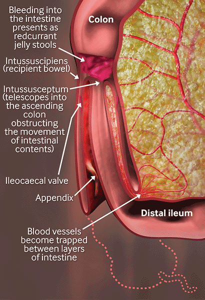
Intussusception is the telescoping of one portion of the intestine (the intussusceptum) into the lumen of the intestine immediately distal to it (the intussuscipiens). The mesentery is dragged alongside of the proximal bowel wall into the distal lumen resulting in obstruction of venous return. Oedema, mucosal bleeding, and increased pressure result. If arterial flow becomes compromised, ischaemia, necrosis, and perforation can occur. Sepsis, shock, and death may eventually occur.[Figure caption and citation for the preceding image starts]: Created by the BMJ Knowledge Centre [Citation ends].
Suggested predisposing factors for idiopathic intussusception include well-developed lymphoid tissue, a thin intestinal wall, a narrow intestinal lumen, poor fixation of the ileocaecal region, viral or bacterial infection, and recent abdominal surgery. These factors are thought to result in mesenteric lymphoid or Peyer's patch hyperplasia, abnormal peristalsis, and bowel-wall oedema.[1]
Ileocolonic intussusception (prolapse of the terminal ileum into the proximal colon) is the most common anatomical location for intussusception to occur, followed by ileoileal and colocolonic.[1]
Use of this content is subject to our disclaimer