Investigations
1st investigations to order
MRI brain
Test
Every child with an equivocal diagnosis of CP should have an MRI of the brain.[86]
Postnatal MRI of the brain is abnormal in up to 80% of patients of CP.[36][37]
MRI is often done in the neonatal intensive care unit at the time of birth in those with an obvious history.[Figure caption and citation for the preceding image starts]: Periventricular leukomalacia. Figure A is a normal 14-year-old female: the curved arrow shows normal lateral ventricle and the straight arrow shows normal white matter. Figure B is a 14-year-old female with CP: the curved arrow shows enlarged ventricle and the straight arrow shows diminished volume of the white matter due to periventricular leukomalaciaCourtesy of Noriko Solomon, Assistant Professor of Radiology, David Geffen School of Medicine at UCLA, Los Angeles [Citation ends].
If the presentation is typical of CP, imaging can be delayed until the child is old enough to have the scan without sedation, typically between ages 5 and 7 years.[37]
Result
periventricular leukomalacia, congenital malformation, stroke or haemorrhage, cystic lesions
Investigations to consider
ultrasound/CT brain
Test
Not as sensitive as MRI in detecting brain abnormalities, but may help in diagnosis and prognosis.[87]
Result
periventricular leukomalacia, congenital malformation, stroke or haemorrhage, cystic lesions
coagulation studies
Test
Consider in patients with hemiplegia who have a high incidence of single-hemisphere infarction.
Result
abnormal in clotting and other haematological disorders
genetic testing
Test
Indicated for patients with dysmorphic features such as abnormal skin creases, low-set ears, lack of a nasal bridge, and hypo- or hypertelorism; or for a suspicion of familial disease.[71] Features may be evident in the neonatal intensive care or may become evident later in childhood.
Result
abnormal in the presence of an underlying genetic disorder
metabolic screen
x-ray of affected joint
Test
Monitoring should start at age 3 years and depends on physical findings (e.g., hip x-rays are needed to monitor for subluxation of hips; spinal x-rays are used to confirm progression in patients with scoliosis, kyphosis, or lordosis). [Figure caption and citation for the preceding image starts]: Dislocated hip in a patient with CPFrom the collection of William L. Oppenheim; used with permission [Citation ends].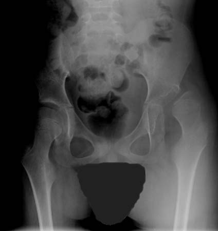
Starting by age 3 years, children with a gross motor function classification system (GMFCS) level I or II need less monitoring; those with level III should be monitored at least every other year during active growth; and those with level IV or V need assessment at least every year during active growth.[90] CanChild: Gross Motor Function Classification System - Expanded & Revised (GMFCS - E&R) Opens in new window
Result
abnormal; dependent on specific complaints or deformity (e.g., equinovarus foot, hip subluxation, spine deformity)
instrumented gait analysis
Test
Gait laboratories offer kinematic and kinetic analysis of gait. Standardised objective tools including computer-motion analysis, electromyography, and force plate recordings are used to identify and quantify features of movement patterns. These data are typically represented in comparison with a database of individuals without disability and provide detailed information on motion and joint forces that cannot be observed clinically.
Result
abnormal movement patterns
Use of this content is subject to our disclaimer