Criteria
MRI classification of juvenile osteochondritis dissecans lesions[33]
Stage description:
Small change of signal without clear margins of fragment
Osteochondral fragment with clear margins but without fluid between fragment and underlying bone
Fluid is visible partially between fragment and underlying bone
Fluid is completely surrounding the fragment, but the fragment is still in situ
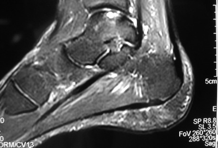
Fragment is completely detached and displaced (loose body).[Figure caption and citation for the preceding image starts]: Sagittal magnetic resonance image (MRI) of the talus, showing an osteochondral lesion on the posterior aspect of the talar domeGupta RK, Kansay R, Aggarwal V, et al. Osteochondritis dessicans of the talus in a 26-year-old woman. BMJ Case Reports 2009; doi:10.1136/bcr.06.2008.0091 [Citation ends].
Transchondral fracture of the talus[34]
Stage description:
Small area of compression of subchondral bone
Partially detached osteochondral fragment
Completely detached osteochondral fragment remaining in the crater
Displaced osteochondral fragment.
Classification of osteochondritis dissecans of the capitellum[35]
Stage description:
Ia-intact/stable: intact articular cartilage/no loss of subchondral stability
Ib-intact/unstable: intact articular cartilage/loss of subchondral stability with impending collapse
II-open/unstable: cartilage fracture/collapse or partial displacement of subchondral bone
III-detached: loose cartilaginous fragment within the joint.
Classification of osteochondritis dissecans severity and stability of the osteochondritis dissecans fragment using arthroscopic and MRI findings[28][29]
Stage description
Stage I
Arthroscopic finding: irregularity and softening of cartilage. No fissure. No definable fragment.
MRI finding: no break, but thickening in articular cartilage.
Stage II
Arthroscopic finding: articular cartilage breached but not displaceable.
MRI finding: articular cartilage breached, low signal rim behind fragment indicating fibrous attachment.
Stage III
Arthroscopic finding: definable fragment, displaceable, but still attached partially by some cartilage (i.e., a flap lesion).
MRI finding: articular cartilage breached with high T2 signal changes behind fragment suggesting fluid behind the lesion.
Stage IV
Arthroscopic finding: loose body and defect of the articular surface.
MRI finding: loose body with defect of articular surface.
Use of this content is subject to our disclaimer