Images and videos
Images
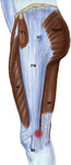
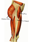


Iliotibial band syndrome
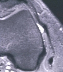
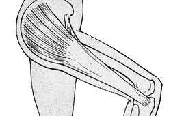
Anatomy of iliotibial band. IT band, iliotibial band; Gluteus max, gluteus maximus; TFL, tensor fascia lata
From the personal collection of Dr J.C. Mak
See this image in context in the following section/s:

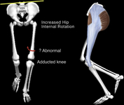
Iliotibial band syndrome
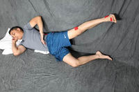
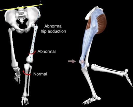
Male runner with iliotibial band syndrome. The femur may or may not exhibit increased internal rotation. The investigators did not report on the mechanism of increased hip internal rotation
Baker RL et al. Iliotibial band syndrome in runners: biomechanical implications and exercise interventions. Phys Med Rehabil Clin N Am. 2016 Feb;27(1):53-77; used with permission
See this image in context in the following section/s:
Iliotibial band syndrome
Injection site for iliotibial band
From the personal collection of Dr J.C. Mak
See this image in context in the following section/s:

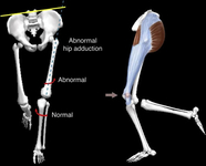
Iliotibial band syndrome
Iliotibial band syndrome. Red mark indicates site of injury: insertion of the iliotibial band into and just proximal to the lateral femoral epicondyle. (BF: biceps femoris; GMAX: gluteus maximus; GT: greater trochanter; ITB: iliotibial band; TFL: tensor fasciae latae; VL: vastus lateralis)
Baker RL et al. Iliotibial band syndrome in runners: biomechanical implications and exercise interventions. Phys Med Rehabil Clin N Am. 2016 Feb;27(1):53-77; used with permission
See this image in context in the following section/s:
Iliotibial band syndrome
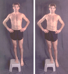
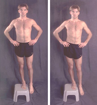
Demonstration of pelvic drop
From the personal collection of Dr J.C. Mak
See this image in context in the following section/s:
Iliotibial band syndrome
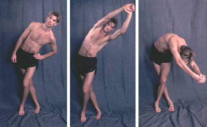
Standing stretch exercise
From the personal collection of Dr J.C. Mak
See this image in context in the following section/s:

Iliotibial band syndrome
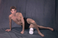
Hip abduction in side-lying position with hip extended for posterior gluteus medius emphasis
Baker RL et al. Iliotibial band syndrome in runners: biomechanical implications and exercise interventions. Phys Med Rehabil Clin N Am. 2016 Feb;27(1):53-77; used with permission
See this image in context in the following section/s:
Iliotibial band syndrome
Female runner with iliotibial band syndrome
Baker RL et al. Iliotibial band syndrome in runners: biomechanical implications and exercise interventions. Phys Med Rehabil Clin N Am. 2016 Feb;27(1):53-77; used with permission
See this image in context in the following section/s:
Iliotibial band syndrome
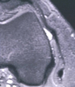
Impingement zone occurring at around 30° of knee flexion
From the personal collection of Dr J.C. Mak
See this image in context in the following section/s:
Iliotibial band syndrome
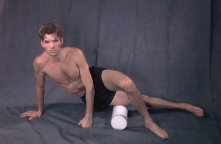
Foam roll exercise
From the personal collection of Dr J.C. Mak
See this image in context in the following section/s:
Videos
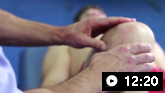 Knee examination
A consultant physician demonstrates knee examination techniques, inspecting the biomechanics of the knee and assessing for effusion, wasting, or hypertension. The physician performs tests for the integrity of the medial and lateral collateral ligaments, damage to the cruciate ligaments (Lackman's test, Drawer test, Pivot shift test), tests of the patella (patella apprehension test, Clarke's test), and tests of the iliotibial band (Ober's test).
Use of this content is subject to our disclaimer