Approach
Characteristic history and examination findings are often sufficient to diagnose the condition.
Presentation
Athletes with ITBS complain of a sharp or burning pain roughly 2 cm superior to the lateral joint line. The pain may radiate proximally or distally. In less severe cases, the pain begins after a reproducible time or distance and subsides quickly upon cessation of activities. With increasing severity, normal walking or sitting with the knee in flexion may become painful.[7] There may also be local oedema and crepitation.
Physical examination
There is usually tenderness on palpation of the iliotibial band (ITB) 2 to 3 cm superior to the lateral joint line. In mild cases results of an examination may be normal, but in severe cases there may be local oedema or crepitation. Noble's, Ober's, and modified Thomas's tests are used in the diagnosis, but are provocative tests used in the physical examination, and not true diagnostic tests. The Noble's test is often positive.
Noble's test: physician applies pressure over the lateral femoral epicondyle while extending the knee from 90° of flexion.[7] Pain occurs when knee is flexed around 30°.
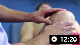
Ober's test: the patient lies down with the unaffected side down and the unaffected hip and knee at a 90° angle. If the ITB is tight, adducting the leg beyond the midline is difficult and the patient may experience pain at the lateral knee. Normal tightness is when the leg can be passively stretched to a position horizontal but not completely adducted to a table. Moderate tightness is when the leg can be passively adducted to horizontal at best. If the leg cannot be passively adducted to horizontal, this is maximal tightness.[7]
Modified Thomas's test: the patient sits on the end of an examining table, rolls back to a supine position, and holds both knees to the chest. The patient holds the knee on the asymptomatic side close to the chest, keeping the hips on the table, and avoiding excessive posterior tilt. The examiner then slowly lowers the affected limb towards the floor.[7] The test is positive if the angle of the femur is below horizontal.
Joint effusion or positive results on a meniscal test will raise suspicion of an intra-articular problem. The Ober's test is recommended to assess tightness of the ITB. The modified Thomas's test is also recommended to evaluate for flexibility deficits in the iliopsoas, rectus femoris, and the tensor fascia lata (TFL)/ITB complex. Hip abductor muscle strength is evaluated in the side-lying position.
Be aware that patients will often compensate for weakness or inhibition of the gluteus medius with substitution of the TFL, the quadratus lumborum muscles, or both. Leg-length discrepancies also contribute to ITBS and are assessed as part of a routine examination.
Imaging
Magnetic resonance imaging (MRI) or ultrasound may be requested if there is doubt about the diagnosis from the physical examination.[30] The result may be normal, or show cystic changes (ultrasound) or poorly defined signal intensity (MRI) changes under the ITB.
MRI of the knee without contrast may be indicated if initial knee x-rays are non-diagnostic (demonstrate normal findings or a joint effusion), or show osteochondral injuries (fracture/osteochondritis dissecans or a loose body), avascular necrosis, or internal derangement (e.g., Segond's fracture, deep lateral femoral notch sign).
A consultant physician demonstrates knee examination techniques, inspecting the biomechanics of the knee and assessing for effusion, wasting, or hypertension. The physician performs tests for the integrity of the medial and lateral collateral ligaments, damage to the cruciate ligaments (Lackman's test, Drawer test, Pivot shift test), tests of the patella (patella apprehension test, Clarke's test), and tests of the iliotibial band (Ober's test).
Use of this content is subject to our disclaimer