Approach
Joint dislocations are usually managed with closed reduction as soon as possible to decrease potential complications including soft tissue injury, articular surface injury, and neurovascular compromise. Reduction usually requires sedation and analgesia.
Patients with abnormal neurological function associated with a joint dislocation should be referred to an orthopaedic specialist.
A period of immobilisation should be followed by active motion exercises and isometric strengthening exercises.
Shoulder dislocation
Once the diagnosis has been confirmed, reduction should be attempted. For a successful outcome, adequate analgesia and sedation are necessary before the reduction procedure is attempted.
There are numerous reduction manoeuvres for shoulder injuries, which are usually performed under local anaesthesia (i.e., intra-articular lidocaine) combined with procedural sedation (e.g., intravenous morphine, midazolam, or etomidate).
[ ![]() ]
Procedural sedation has the added advantage of reducing spasm in the muscles of the rotator cuff. The choice for sedation depends on the treating physician and must be accompanied by continuous monitoring of the patient with capnography and pulse oximetry, as well as frequent blood pressure measurements. Monitoring should begin before any medicines are administered and continue until the patient is fully awake. Multiple studies have shown that local anaesthesia on its own is equivalent to intravenous sedation.[64][65][66] However, local anaethesia on its own should be reserved for patients with contraindications to procedural sedation.
]
Procedural sedation has the added advantage of reducing spasm in the muscles of the rotator cuff. The choice for sedation depends on the treating physician and must be accompanied by continuous monitoring of the patient with capnography and pulse oximetry, as well as frequent blood pressure measurements. Monitoring should begin before any medicines are administered and continue until the patient is fully awake. Multiple studies have shown that local anaesthesia on its own is equivalent to intravenous sedation.[64][65][66] However, local anaethesia on its own should be reserved for patients with contraindications to procedural sedation.
Each reduction method works by abduction and external rotation to disengage the humeral head from the glenoid, with axial traction to reduce it. The following are examples of reduction manoeuvres for anterior dislocations.
Traction-countertraction method
The patient is placed supine on the bed. A sheet is looped around the axilla with one free end on the chest and the other underneath the back. The 2 ends should be of even length. An assistant uses these free ends to apply countertraction. Then the practitioner abducts the arm to 90° and flexes the elbow to 90°. With the forearm, slow longitudinal traction is then applied to the affected extremity. [Figure caption and citation for the preceding image starts]: Traction-countertraction method: with patient supine on the bed, a sheet is looped around the axilla with one free end on the chest and the other underneath the back; the 2 ends should be of even length and are used by an assistant to apply countertraction. The practitioner abducts the arm to 90° and flexes the elbow to 90°; the forearm is used to apply slow longitudinal traction to the affected extremityPersonal collection of Dr Paul Novakovich [Citation ends].

Modified Milch's technique
The modified Milch's technique has been reported to be effective in 100% of dislocations without the use of sedation.[67] The patient is positioned supine on a bed with the head of the bed elevated approximately 20° to 30°. The arm is slowly abducted and externally rotated without application of longitudinal traction. The practitioner pauses in the case of pain or resistance. Once the arm has reached a position of 90° abduction and 90° external rotation, the shoulder should spontaneously reduce. If not, the humeral head can be palpated in the axilla, and superolateral pressure can then be applied using the thumb and index finger to help guide the humeral head back into the glenoid.[55][67][Figure caption and citation for the preceding image starts]: Milch's technique for shoulder reduction, part 1: patient is positioned supine on a bed with the head of the bed elevated about 20° to 30°, then the arm is slowly abducted and externally rotated without application of longitudinal traction (in case of pain or resistance, the practitioner pauses)Personal collection of Dr Paul Novakovich [Citation ends].
 [Figure caption and citation for the preceding image starts]: Milch's technique for shoulder reduction, part 2: once the arm has reached a position of 90° abduction and 90° external rotation, the shoulder dislocation should spontaneously reduce; if not, the humeral head can be palpated in the axilla and superolateral pressure can then be applied using the thumb and index finger to help guide the humeral head back into the glenoidPersonal collection of Dr Paul Novakovich [Citation ends].
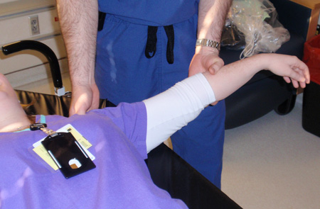
Stimson's method or scapular manipulation technique
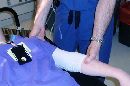
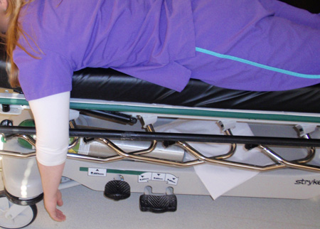
This method is reported to be effective in 96% of shoulder dislocations.[68] The patient is positioned prone on the stretcher with the affected shoulder slightly off the stretcher. The arm is placed perpendicular to the floor (90° forward flexion) with the stretcher high enough to keep the hand from resting on the floor. Weights of 2.3 to 4.5 kg or 1-L bottles of sterile water are wrapped around the wrist using stockinette and hung high enough to not touch the floor. Reduction should occur within 10 to 20 minutes. Reduction can be facilitated by external rotation, or application of the scapular manipulation technique (where one hand is placed on the superolateral border of the scapula and the other hand on the inferomedial border of the scapula, and pressure is applied to rotate the superior border laterally and the inferior border medially).[55][68][Figure caption and citation for the preceding image starts]: Stimson's method: patient is positioned prone on the stretcher with the affected shoulder slightly off the stretcher; arm is placed perpendicular to the floor (90° forward flexion) with the stretcher high enough to keep the hand from resting on the floor, then weights of 2.3 to 4.5 kg or 1-L bottles of sterile water are wrapped around the wrist using stockinette and hung high enough to not touch the floorPersonal collection of Dr Paul Novakovich [Citation ends].
 [Figure caption and citation for the preceding image starts]: Scapular manipulation technique: one hand is placed on the superolateral border of the scapula with the other hand on the inferomedial border of the scapula, and pressure is applied to rotate the superior border laterally and the inferior border mediallyPersonal collection of Dr Paul Novakovich [Citation ends].
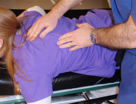
Sitting scapular manipulation technique
The patient sits upright with an assistant applying scapular pressure using the scapular manipulation technique. The practitioner then flexes the affected arm to 90°, placing one hand on the clavicle and the other on the wrist. Gentle traction is applied to the arm with pressure applied to the clavicle. Simultaneously, the assistant performs the scapular manipulation.
Spaso technique
This is reported to be effective in 88% of shoulder dislocations.[69] The patient is positioned supine on the bed. The affected shoulder is slowly forward flexed to 90°. Gentle longitudinal traction is applied while the shoulder is slowly externally rotated. Care must be taken to keep the medial border of the scapula firmly pressed against the bed.
External rotation method
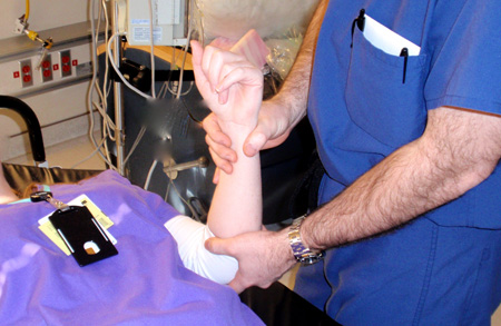
This was initially described by Leidelmeyer and is reported to be successful in 78% of patients.[66] The patient is positioned supine on the bed and the affected extremity is gently adducted until it is parallel to the long axis of the body. The elbow is then flexed to 90°. By applying gentle pressure to the wrist, the practitioner slowly externally rotates the arm, taking time to allow spasms and contractions to pass. Finally, the arm is externally rotated to 90° (i.e., perpendicular to the long axis of the body). After approximately 5 minutes, the shoulder should reduce. [Figure caption and citation for the preceding image starts]: External rotation method for shoulder reduction, part 1: patient is positioned supine, and affected extremity is gently adducted until it is parallel to the long axis of the body; elbow is then flexed to 90°Personal collection of Dr Paul Novakovich [Citation ends].
 [Figure caption and citation for the preceding image starts]: External rotation method for shoulder reduction, part 2: by applying gentle pressure to the wrist, the practitioner slowly externally rotates the arm, taking time to allow spasms and contractions to pass; finally, the arm is externally rotated to 90° (i.e., perpendicular to the long axis of the body); shoulder dislocation should reduce after about 5 minutesPersonal collection of Dr Paul Novakovich [Citation ends].
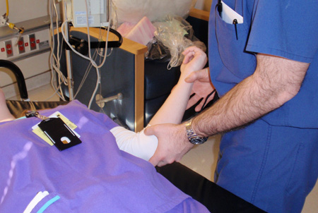
Kocher's method
This method is more complex than other methods and may be associated with complications (e.g., fractures, brachial plexus injury and vascular injury). It uses in-line traction of the arm while abducted to 45°. While traction is maintained, the arm is externally rotated and the elbow is brought across the chest to the mid-line. The arm is then internally rotated until the patient's hand touches the shoulder. [Figure caption and citation for the preceding image starts]: Kocher's method of shoulder reduction, part 1: in-line traction of the arm while abducted to 45°; while traction is maintained, arm is externally rotated and elbow is brought across the chest to the mid-linePersonal collection of Dr Paul Novakovich [Citation ends].
 [Figure caption and citation for the preceding image starts]: Kocher's method of shoulder reduction, part 2: arm is internally rotated until the patient's hand touches the shoulderPersonal collection of Dr Paul Novakovich. [Citation ends].
FARES (Fast Reliable and Safe) method
The physician holds the hand of the patient at the patient's side with the elbow extended and in neutral rotation. Longitudinal traction is applied and the arm is moved into abduction (no countertraction needed). As the arm is abducted, short vertical oscillations are performed to relax the musculature. As the arm passes 90° of abduction, the arm is then externally rotated, and at 120° of arm abduction, the arm typically reduces. When compared with the Kocher and traction-countertraction methods, this method is faster and requires less sedation.[70]
Irrespective of the technique used, the physician should feel a distinct clunk as the shoulder reduces. The arm should be immobilised and placed in a sling or a sling and swathe. Anteroposterior (AP) and lateral x-rays should be obtained to confirm reduction of the humeral head and to ensure that no iatrogenic fractures have occurred during the reduction.[71]
Once the patient is alert, it is important to perform a neurological examination, with emphasis on the axillary, radial, ulnar, and median nerves. The vascular status of the hand should also be re-assessed to ensure that the axillary or brachial artery has not been injured during the reduction.
The patient should wear the sling for approximately 3 weeks. In subsequent weeks, active-assisted range of motion and isometric strengthening exercises should be advised. Generally, by week 12, limited return to sporting activities is permitted, followed by full return to sporting activities as tolerated by week 16. [Figure caption and citation for the preceding image starts]: Normal axillary x-ray view of a reduced shoulder dislocation, showing congruency of the glenohumeral jointPersonal collection of Dr Paul Novakovich [Citation ends].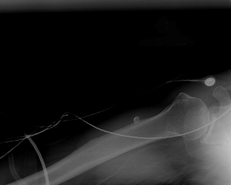
Surgical referral
Patients under 25 years of age should be referred to an orthopaedic surgeon for assessment, as this age group is at significant risk for recurrence. Long-term data support primary stabilisation via anatomical Bankart's repair (over simple arthroscopic lavage or non-operative treatment) for young, high-risk patients with a first-time shoulder dislocation.[72][73] One systematic review found that patients, particularly active men in their 20s and 30s, undergoing treatment for a first-time anterior shoulder dislocation with a surgical stabilisation procedure, can be expected to experience significantly lower rates of recurrent instability and a significantly decreased need for a future stabilisation procedure when compared with patients treated non-operatively.[74]
The Latarjet's procedure is a commonly used approach for managing chronic and recurrent anterior shoulder dislocation, especially in the presence of bone loss.[75] It involves transplant of the coracoid process to the scapular neck and has demonstrated excellent long-term clinical outcomes and return to sport rate.[76] It may be more effective than Bankart's repair for recurrent instability of the shoulder.[77] Both open and arthroscopic Latarjet's procedures result in significantly improved function and outcome in patients with anterior shoulder instability.[78] However, the Latarjet's procedure has been associated with a complication rate of 15% to 30%; specific complications include graft-related issues (11.7%), hardware-related complications (6.5%), nerve injuries (0.7% to 4%), recurrent instability (8%), and revision (5%).[75]
Finger dislocation
The goal of treatment for finger dislocations is to restore joint congruity by means of closed reductions. Certain situations can make congruent reduction difficult. These include volar plate entrapment, volar dislocations, and fracture dislocations.
Reduction of finger dislocation often requires the use of a local anaesthetic, typically lidocaine 1%. A neurovascular examination of the digit is essential before reduction is performed because the local anaesthetic may cause complete hypoaesthesia of the finger. Two sets of nerves run on the radial and ulnar side of each digit. Generally, the dorsal nerves lie on the 10-o'clock and 2-o'clock positions, while the palmar digital nerves lie on the 8-o'clock and 4-o'clock positions. Infiltration of lidocaine in these quadrants results in effective anaesthesia for reducing a dislocated finger.
Dorsal proximal interphalangeal (PIP) and distal interphalangeal (DIP) dislocations
The first step in reduction is to recreate the injury by hyper-extending the PIP or DIP. This should be followed by light axial traction applied to the finger with pressure applied to the base of the dislocated digit until the joint is relocated. Occasionally, these joints will not reduce because of entrapment of the volar plate, and consultation with a hand surgeon will be required. If the joint is stable, buddy taping to an adjacent digit or placement of a splint in slight flexion is an appropriate measure. Neutral splinting for dorsal PIP dislocations can also be used and is reported to avoid post-splinting flexion contractures.[79] Post-reduction x-rays should be obtained to confirm congruency of the joint and to ensure that there are no associated fractures.[15]
Volar DIP and PIP dislocations
These are more likely to be unstable, but the goals of reduction are the same as for dorsal dislocations. The finger should be flexed with mild axial traction applied to the digit. The physician should then apply pressure to the base of the digit until reduction is complete. Post-reduction x-rays should be obtained to confirm congruence of the joint and to ensure that there are no associated fractures. The finger should be placed in an extension splint immobilising the smallest number of joints possible. If concentric reduction is not possible because of soft tissue entrapment, consultation with a hand surgeon is warranted.
Metacarpophalangeal (MCP) dislocation
With simple dislocations, the finger is usually held in extension, and there is some contact between the joint surfaces. The wrist should be flexed to relax the flexor tendons, and the affected digit should then be hyper-extended. The physician should then apply a volar-directed pressure to the dorsum of the affected digit. It is paramount that excessive traction not be applied, as a simple dislocation can be converted into a complex MCP dislocation with significant soft tissue entrapment. If this occurs, the joint will often become irreducible and require operative treatment.[24][80]
Simple dislocations can be buddy taped, while fracture dislocations require immobilisation in a splint. Post-reduction x-rays should be obtained to confirm congruence of the joint and to ensure that there are no associated fractures. Following reduction, the physician should ensure adequate perfusion to the finger by assessing capillary refill. Post-reduction, patients should begin protected range of motion as pain permits. In treating finger dislocations, instituting early motion and providing stability must be balanced.
Patellar dislocation
Patellar dislocation often presents to the emergency department or to the clinic having already spontaneously reduced.
In the patient presenting with an acute patellar dislocation, a reduction should be performed with the goal being the concentric reduction of the patella into the femoral notch. Patellar dislocations have been reported to be accompanied by intra-articular lesions in 5% to 71% cases.[16][56][57] In these cases, orthopaedic consultation is warranted, as open reduction surgery may be required.
For a successful outcome, adequate analgesia and sedation is necessary before the reduction procedure is attempted. Lateral dislocation is easily managed using local anaesthesia (i.e., intra-articular lidocaine) combined with procedural sedation (e.g., intravenous morphine, midazolam, or etomidate). Procedural sedation has the added advantage of reducing muscle spasm. The choice for sedation depends on the treating physician and must be accompanied by continuous monitoring of the patient with capnography and pulse oximetry, as well as frequent blood pressure measurements. Monitoring should begin before any medicines are administered and continue until the patient is fully awake.
Following adequate analgesia and sedation, the patient should be placed either supine or in a seated position. The affected knee should be flexed to decrease the tension on the quadriceps muscle. The physician should apply a medial-directed force to the lateral aspect of the patella while slowly extending the leg. A palpable clunk should confirm reduction of the patella. Upon successful reduction, the affected extremity should be placed in a knee immobiliser and the patient advised to bear weight on the joint as tolerated.
Merchant, AP, and lateral knee plain x-rays should be ordered to ensure that the patella is reduced. The x-rays should be closely examined for evidence of any osteochondral defects that may have been created during the reduction. Post-reduction, patients should begin protected range of motion as pain permits.
Surgery versus non-operative approach
A Cochrane review concluded that large multicentre clinical trials are needed to determine whether a surgical or non-operative approach is preferred for the management of patellar dislocation.[81] A systematic review of overlapping meta-analyses found that operative treatment of first-time patellar dislocations results in a lower recurrence rate but no improvement in functional outcome scores compared with non-operative management.[82]
Medial patellofemoral ligament reconstruction for patellofemoral instability was associated with a high rate of success in one systematic review of the literature.[83] Complications were, however, common (complication rate 26.1%).
Elbow dislocation
For a successful outcome, adequate analgesia and sedation are necessary before the reduction procedure is attempted.
The reduction is usually performed using local anaesthesia (i.e., intra-articular lidocaine) combined with procedural sedation (e.g., intravenous morphine, midazolam, or etomidate). Procedural sedation has the added advantage of reducing muscle spasm. The choice for sedation depends on the treating physician and must be accompanied by continuous monitoring of the patient with capnography and pulse oximetry, as well as frequent blood pressure measurements. Monitoring should begin before any medicines are administered and continue until the patient is fully awake.
The patient should be supine on the bed with the physician positioned on the affected side with an assistant close to the head of the bed. In young children, management of radial head subluxation/dislocation (nursemaid's elbow) may be more effective and less painful when performed with the arm in pronation as opposed to supination.[84]
[ ![]() ]
The arm should be initially extended to 30° flexion. The overall gross alignment of the elbow is then manipulated so that the olecranon appears centred between the medial and lateral condyle of the humerus. The forearm is then slowly flexed to approximately 90° with the physician providing longitudinal traction to the forearm while the assistant provides countertraction to the patient's humerus. The arm is then flexed even further with direct downward pressure applied to the olecranon.[44]
]
The arm should be initially extended to 30° flexion. The overall gross alignment of the elbow is then manipulated so that the olecranon appears centred between the medial and lateral condyle of the humerus. The forearm is then slowly flexed to approximately 90° with the physician providing longitudinal traction to the forearm while the assistant provides countertraction to the patient's humerus. The arm is then flexed even further with direct downward pressure applied to the olecranon.[44]
If reduction is successful, the physician should feel an audible clunk as the elbow is reduced. It is important not to flex the arm forcefully if there is significant resistance because the coronoid process is typically perched on the distal humerus. Forceful flexion without adequate traction can cause a fracture of this structure, which will result in future instability. Upon reduction, the arm is placed in a posterior splint at 90° flexion with neutral rotation of the forearm.[44] An AP and lateral plain film radiograph of the elbow should be obtained to ensure that the joint is concentrically reduced.
Closed reduction of posterolateral dislocation of the elbow
Once the patient is alert, it is important to perform a neurological examination, with emphasis on the radial, ulnar, and median nerves. The vascular status of the hand should also be re-assessed to ensure that the brachial artery has not been injured during the reduction. Several studies have shown better outcomes with early mobilisation than with immobilisation in patients with simple dislocations. Patients should initially be splinted in a posterior splint for comfort with instructions to begin mobilisation when pain permits. Immobilisation should last no longer than 2 weeks.[33][85][86][87][88][Figure caption and citation for the preceding image starts]: Anteroposterior x-ray view of a reduced elbow dislocationPersonal collection of Dr Paul Novakovich [Citation ends].
Hip dislocation
Early reduction and avoidance of complications (e.g., osteonecrosis) is the immediate goal in the setting of an acute hip dislocation.
Every effort should be made to obtain reduction of the dislocated hip within 6 hours from injury, via closed or open reduction techniques to maximise functional recovery.[4] Urgent reduction of the femoral head into the acetabulum is indicated in almost all cases. The tenuous blood supply to the femoral head is easily compromised with a dislocation, and the incidence of osteonecrosis has been shown to increase if reduction is delayed. Unless an associated hip or femoral neck fracture is known to exist, a closed reduction under sedation or anaesthesia can be attempted in the accident and emergency department.[89]
There are several described techniques for reduction of both anterior and posterior hip dislocations. Hip reduction can be attempted using in-line traction, with the patient lying supine, followed by applying a force opposing the vector of the initial injury. Initially, the traction should be applied in a steady manner to overcome muscular spasms.
Allis's method:
Traction is applied in line with the deformity. The patient is placed supine, with the surgeon standing above the patient on the stretcher. Initially, the surgeon applies in-line traction, while the assistant applies counter-traction, stabilising the pelvis. While increasing the traction force, the surgeon slowly increases the degree of flexion to approximately 70°. Gentle rotational motions of the hip and slight adduction will often help the femoral head clear the lip of the acetabulum. A lateral force to the proximal thigh may assist in reduction. An audible 'clunk' is a sign of a successful closed reduction.[90]
Stimson's gravity technique:
The patient is placed prone on the stretcher, with the affected leg hanging off the side of the stretcher. This brings the extremity into a position of 90° of both hip and knee flexion. In this position, the assistant immobilises the pelvis and the surgeon applies an anteriorly directed force on the proximal calf. Gentle rotation of the limb may assist in reduction.[90]
Hip immobilisation is difficult. Patients usually do well with assisted ambulation using crutches, and bear weight as tolerated. Crutches should be used until the patient can walk relatively pain-free, and the knee immobiliser should be kept in place until strength improves and symptoms abate. Moderate quadriceps strengthening should begin when the patient is comfortable.
Use of this content is subject to our disclaimer