Investigations
1st investigations to order
plain x-rays of the shoulder
Test
An anteroposterior (AP) x-ray view of the shoulder should be taken with internal and external humeral rotation.[59]
These should be accompanied by axillary lateral and/or scapular Y views to confirm diagnosis. Each of these x-ray views is 92% sensitive for acute shoulder dislocation.[60]
On a scapular Y view, the humeral head lies anteriorly to the Y in anterior dislocations, and posteriorly to the Y in posterior dislocations. In standard AP views, the humeral head rests anteroinferiorly to the coracoid in anterior dislocations. However, in posterior dislocations, the humerus can appear to be reduced. Therefore, axillary or scapular Y views are essential for accurate diagnosis, as up to 79% of posterior shoulder dislocations are initially misdiagnosed. Axillary views can correctly identify posterior dislocations in 100% of patients when combined with AP views of the shoulder.[61][62]
An axillary oblique or modified axillary (Velpeau or West Point) view may also be used to confirm diagnosis.
A possible fracture of the proximal humerus should be excluded, as attempts at reduction could further displace this fracture. [Figure caption and citation for the preceding image starts]: Scapular Y x-ray view showing an anterior fracture dislocation of the shoulder and fracture of the greater tuberosityPersonal collection of Dr Paul Novakovich [Citation ends]. [Figure caption and citation for the preceding image starts]: Anteroposterior x-ray view of a shoulder showing an anteroinferior dislocationPersonal collection of Dr Paul Novakovich [Citation ends].
 [Figure caption and citation for the preceding image starts]: Anteroposterior x-ray view of a shoulder showing a missed posterior dislocation: the glenohumeral joint appears reducedPersonal collection of Dr Paul Novakovich [Citation ends].
 [Figure caption and citation for the preceding image starts]: Axillary lateral of a shoulder with a missed posterior dislocation: humeral head clearly is not reduced and is locked on the posterior rim of the glenoidPersonal collection of Dr Paul Novakovich [Citation ends].
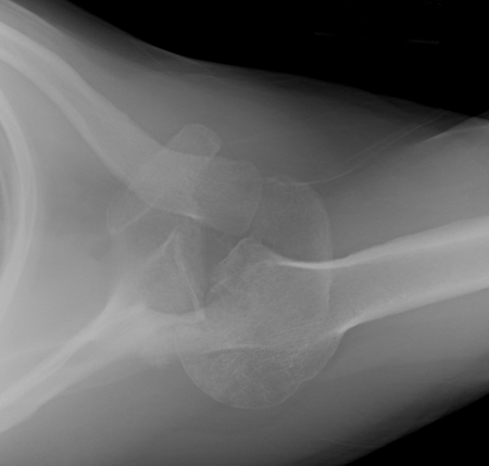
Result
incongruity, subluxation, or loss of reduction of the glenohumeral joint
plain x-rays of the finger
Test
Imaging is usually not necessary in the acute management of simple finger dislocations.
However, anteroposterior, oblique, and lateral views of the affected joint are mandatory in evaluating a hand injury to exclude fracture and/or dislocation.
X-rays should be closely inspected for associated fractures and avulsions, which may indicate ligament or tendon damage.
[Figure caption and citation for the preceding image starts]: X-ray showing dislocation of the proximal interphalangeal joint, left index fingerHellerhoff, CC BY-SA 3.0 via Wikimedia Commons [Citation ends].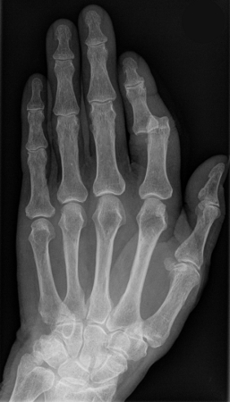
Result
incongruity of the proximal interphalangeal, distal interphalangeal, and/or metacarpophalangeal joint or a fracture
plain x-rays of the knee
Test
Anteroposterior and lateral views of the knee are required. However, it is not uncommon for patella dislocations to reduce spontaneously during examination or following leg extension.
Dislocation can be confirmed on a Merchant or sunrise (infra-patellar) view, which demonstrates the medial facet of the patella resting on the lateral trochlea of the femur.
Images should also be inspected closely for evidence of osteochondral lesions.
[Figure caption and citation for the preceding image starts]: Left knee radiograph demonstrating lateral patella dislocationYerimah G, et al. BMJ Case Rep. 2013 May 2:2013:bcr2013009832; used with permission [Citation ends].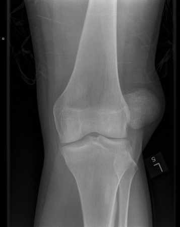
Result
incongruity of the patellofemoral joint, or associated osteochondral fracture
plain x-rays of the elbow
Test
In a posterior dislocation, standard AP and lateral views of the elbow joint show the radius and ulna lying posterior to the distal humerus.
The radial head should always be in line with the capitellum, and the olecranon should rest in the trochlea on a standard lateral view.
AP and lateral x-ray views of the forearm are also necessary to exclude associated forearm fractures (e.g., Monteggia fracture).
A radial head/capitellum view may be used to discern radial head and coronoid fractures. [Figure caption and citation for the preceding image starts]: Lateral x-ray view of a posterolateral elbow dislocationPersonal collection of Dr Paul Novakovich [Citation ends].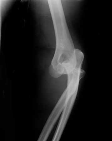 [Figure caption and citation for the preceding image starts]: Anteroposterior x-ray view of an elbow dislocationPersonal collection of Dr Paul Novakovich [Citation ends].
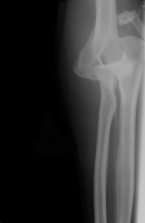
Result
incongruity of the radiocapitellar joint and the humeroulnar joint
plain x-rays of the pelvis
Test
Pelvic x-rays (Judet views, and inlet and outlet views) are routinely obtained to assess for possible concomitant fractures to the pelvis. [Figure caption and citation for the preceding image starts]: X-ray showing bilateral hip posterior dislocationFan KY, et al. BMJ Case Rep. 2015 Mar 25:2015:bcr2014204031; used with permission [Citation ends].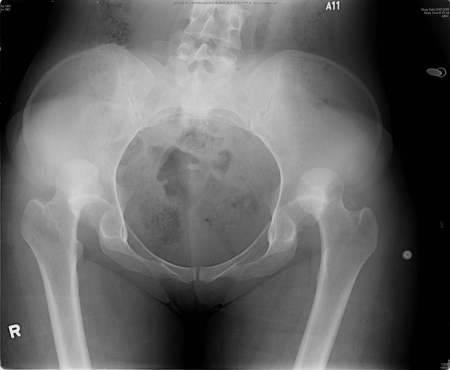
Result
incongruity of the hip joint (anterior or posterior), or fracture of the femoral neck or pelvis
Investigations to consider
MRI scan of the knee
Test
Provides excellent visualisation of soft tissue anatomy and should be ordered if any associated injuries are suspected.
Useful in evaluating concomitant knee injuries including ligamentous and osteochondral lesions.
Result
incongruity of the patellofemoral joint, or associated osteochondral fracture
MRI scan of the shoulder
Test
Provides excellent visualisation of soft tissue anatomy and should be ordered if any associated injuries are suspected.
Can be useful to exclude concomitant rotator cuff tears, which are not uncommon.
Result
rotator cuff tears
CT scan of the elbow
Test
Can further delineate fractures (e.g., of the radial head and coronoid process) if plain film radiography is insufficient to confirm diagnosis.
Result
incongruity of the radiocapitellar joint and the humeroulnar joint
CT scan of the pelvis
Test
Can identify specific fracture patterns, as well as smaller bony lesions (loose bodies) that may prevent a closed reduction and mandate an open reduction in the operating room.[4]
Result
fracture of the femoral neck or pelvis; presence of loose bodies
arteriogram of the knee or shoulder
Test
If a vascular injury is of concern based on the physical examination, an arteriogram may be required.
Allows radiographic evaluation of the injured vessel by demonstrating interruption of arterial blood flow from the proximal to the distal end of the affected vessel.
Result
disruption of integrity of the popliteal artery around the knee, axillary artery in the shoulder, and brachial artery in the arm, with concomitant vascular injury
Emerging tests
ultrasound of the shoulder
Test
Point-of-care ultrasound (POCUS) has been suggested as an additional diagnostic tool for shoulder dislocation. It has the potential to reduce time to diagnosis and reduction, as well as radiation exposure and healthcare costs. One systematic review found that POCUS had 100% sensitivity and specificity for the identification of shoulder dislocations and reductions, and 96.8% sensitivity and 99.7% specificity for the detection of associated fractures.[63]
Result
humeral head displaced anterior or posterior to the glenoid; an anechoic or hypoechoic disruption in the normal contour of the (hyperechoic) bone is seen if coexistent fracture is present
Use of this content is subject to our disclaimer