Investigations
1st investigations to order
optical coherence tomography
Test
Should be ordered for all patients at baseline; for patients with exudation, haemorrhages, or microaneurysms in the macular area; and for patients with unexplained visual loss.[Figure caption and citation for the preceding image starts]: Optical coherence tomography in macular oedema: loss of central foveal depression (yellow arrow), accumulation of fluid within cystoid spaces at fovea (red arrow)Courtesy of Moorfields Photographic Archive; used with permission [Citation ends].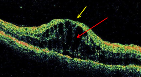 [Figure caption and citation for the preceding image starts]: Optical coherence tomogram of normal eye: normal foveal depression at centre of macula (yellow arrow), inner retina (towards centre of eye; red arrow), outer retina (further from centre of eye; blue arrow)Courtesy of Moorfields Photographic Archive; used with permission [Citation ends].
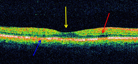 [Figure caption and citation for the preceding image starts]: Optical coherence tomography in vitreomacular traction: loss of foveal depression with traction on fovea (in direction of yellow arrow)Courtesy of Moorfields Photographic Archive; used with permission [Citation ends].
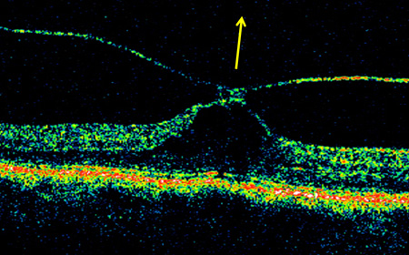
Result
May be used to diagnose: macular oedema (intraretinal cystic spaces, increase in centrepoint thickness, increase in central subfield thickness; also, determining whether centre-involving or non-centre-involving), macular ischaemia (disorganisation of the retinal inner layers, foveal ganglion cell loss, outer retinal atrophy), vitreomacular traction (posterior hyaloid thickening, tractional retinoschisis), vitreous attachment to neovascularisation of retina or optic disc; may also be used to monitor response to treatment of macular oedema
fundus photography/wide-field fundus photography
Test
Should be ordered for all patients at baseline, and when there is a change in fundus appearance. [Figure caption and citation for the preceding image starts]: Normal retina left eye: optic disc (white square), macula (white circle), arteriole (red arrow), venule (blue arrow)Courtesy of Moorfields Photographic Archive; used with permission [Citation ends].
Result
may be used to assess: retinopathy severity; retinopathy progression
Investigations to consider
fluorescein angiography/wide-field fluorescein angiography
Test
Indicated in some patients with diabetic maculopathy, and some patients with severe non-proliferative/proliferative retinopathy.[16]
[Figure caption and citation for the preceding image starts]: Fluorescein angiogram in mid-venous phase in diabetic retinopathy with microaneurysms only: microaneurysm (red arrow), optic disc (yellow arrow), macula (white circle)Courtesy of Moorfields Photographic Archive; used with permission [Citation ends].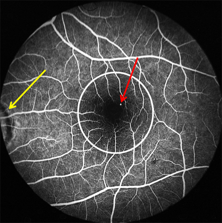 [Figure caption and citation for the preceding image starts]: Fluorescein angiogram of non-proliferative diabetic retinopathy: microaneurysms (red arrow), intraretinal microvascular abnormalities (blue arrow)Courtesy of Moorfields Photographic Archive; used with permission [Citation ends].
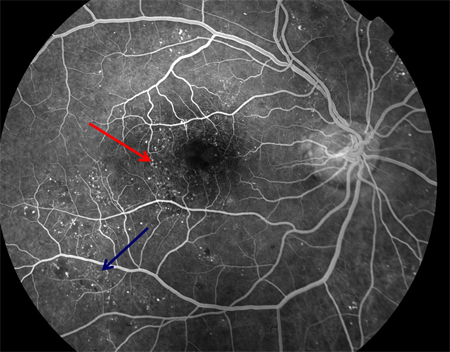 [Figure caption and citation for the preceding image starts]: Fluorescein angiogram in proliferative diabetic retinopathy with macular ischaemia: macular ischaemia (green circle), capillary non-perfusion (white arrow), optic disc new vessels (red arrow), venous beading (blue arrow)Courtesy of Moorfields Photographic Archive; used with permission [Citation ends].
 [Figure caption and citation for the preceding image starts]: Fluorescein angiography in proliferative diabetic retinopathy. Vascular component of fibrovascular proliferation (red arrows), capillary non-perfusion (yellow arrow), laser burns (green circle)Courtesy of Moorfields Photographic Archive; used with permission [Citation ends].
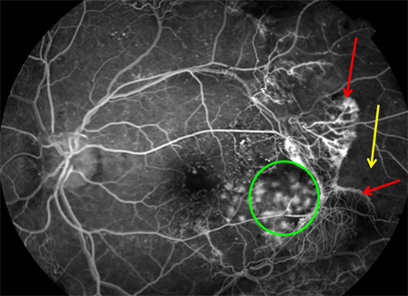 [Figure caption and citation for the preceding image starts]: Fluorescein angiogram in proliferative diabetic retinopathy: new vessels on the optic disc (red arrow), capillary non-perfusion (red rectangle), microaneurysms (green circle), venous beading (blue arrow), intraretinal microvascular abnormalities (yellow arrow)Courtesy of Moorfields Photographic Archive; used with permission [Citation ends].
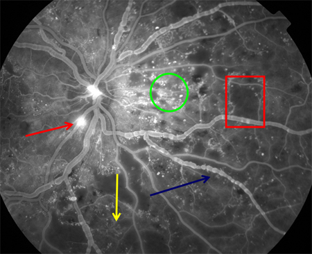 [Figure caption and citation for the preceding image starts]: Fluorescein angiography in proliferative diabetic retinopathy: new vessels elsewhere (red circle), capillary non-perfusion (white arrow), pan-retinal laser burns (blue arrow)Courtesy of Moorfields Photographic Archive; used with permission [Citation ends].
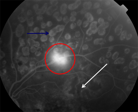
Result
defines macular leakage (versus optical coherence tomography definition of thickness); quantifies macular ischaemia; determines extent of capillary non-perfusion; identifies new vessels; determines adequacy of pan-retinal photocoagulation
optical coherence tomography angiography
Test
Indicated in patients with suspected macular ischaemia.[71]
[Figure caption and citation for the preceding image starts]: Optical coherence tomography in macular oedema: loss of central foveal depression (yellow arrow), accumulation of fluid within cystoid spaces at fovea (red arrow)Courtesy of Moorfields Photographic Archive; used with permission [Citation ends].[Figure caption and citation for the preceding image starts]: Optical coherence tomogram of normal eye: normal foveal depression at centre of macula (yellow arrow), inner retina (towards centre of eye; red arrow), outer retina (further from centre of eye; blue arrow)Courtesy of Moorfields Photographic Archive; used with permission [Citation ends].[Figure caption and citation for the preceding image starts]: Optical coherence tomography in vitreomacular traction: loss of foveal depression with traction on fovea (in direction of yellow arrow)Courtesy of Moorfields Photographic Archive; used with permission [Citation ends].
Result
identifies vascular abnormalities in deep and superficial capillary complexes
B-scan ultrasonography
Test
Should be ordered in patients in whom media opacity (e.g., vitreous haemorrhage) prevents visualisation of the fundus.
Result
identifies macular traction retinal detachment, and other major structural derangements of the retina
Use of this content is subject to our disclaimer