Approach
The diagnosis of psoriatic arthritis (PsA) is made clinically based on history and physical examination.
There are a number of screening questionnaires to identify PsA. The Psoriatic and Arthritic Questionnaire (PAQ), Psoriatic Arthritis Screening and Evaluation (PASE), Psoriasis Epidemiology Screening Tool (PEST), and electronic Psoriasis and Arthritis Screening Questionnaire (ePASQ) were developed to identify PsA in patients with psoriasis, while others were developed to identify PsA in any setting, such as the Toronto Psoriatic Arthritis Screen (ToPAS), and the Toronto Psoriatic Arthritis Screen Version 2 (ToPAS2).[26][27][28][29][30][31]
Laboratory and radiological investigations are obtained either to stage the severity of the disease or to exclude other diagnoses.
Recognition of inflammatory joint symptoms is the first step. Symptoms that suggest PsA in a patient with psoriasis include:
prolonged morning stiffness in joints (lasting >30 minutes)
morning first-step foot pain, and
tender/swollen joints.
These symptoms may be similar to those of early rheumatoid arthritis (RA). These two types of inflammatory arthritis can often be distinguished clinically by the pattern of joint involvement, and the presence of psoriasis along with nail changes typical for psoriasis. [Figure caption and citation for the preceding image starts]: Psoriatic arthritisFrom the collection of Dr Tsu-Yi Chuang; used with permission [Citation ends].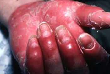
History
PsA presents most commonly in people with an established case of psoriasis.[32] However, not all of these patients are aware that they have psoriasis, so the history of evolution and pattern of joint symptoms should lead to questioning the possibility of psoriatic disease in the patient and/or family.
To begin with it is important to define the nature of the musculoskeletal complaint.
Are inflammatory joint symptoms present (characterised by prolonged morning stiffness >30 minutes, improvement with use, and recurrence with prolonged rest)?
Are the symptoms limited, asymmetrical, monoarticular, or oligoarticular? PsA frequently presents with a pattern of monoarticular or oligoarticular joint involvement. Polyarticular joint involvement, or dactylitis, is a high-risk presentation and is associated with significant progression and poor outcome.
Is there involvement of individual digits (ray pattern rather than row pattern, as is seen in symmetrical RA)?
Are the lower extremity symptoms worse than upper? Early PsA may involve just a few lower extremity joints such as the knees, ankles, or toes.
Are there symptoms suggestive of enthesitis (heel pain, elbow pain, or lateral hip pain related to entheseal inflammation) at bony tendon or ligament attachments?
Does the patient have inflammatory back pain along with spinal stiffness (especially cervical spine)?
Is there a personal or family history of psoriasis?
Does the patient have a personal history of scalp or nail problems (suggesting underlying psoriasis)?
Does the patient have extra-articular manifestations, such as uveitis or inflammatory bowel disease?
Physical examination
Evidence of psoriasis should be sought. Lesions are red, inflamed, silvery-white, scaly, circumscribed papules and plaques commonly found on elbows, knees, extensor limbs, and the scalp.[Figure caption and citation for the preceding image starts]: Psoriasis plaque: legsFrom the collection of Dr Tsu-Yi Chuang; used with permission [Citation ends].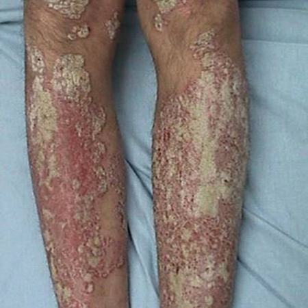 [Figure caption and citation for the preceding image starts]: Psoriasis plaque: scalpFrom the collection of Dr Tsu-Yi Chuang; used with permission [Citation ends].
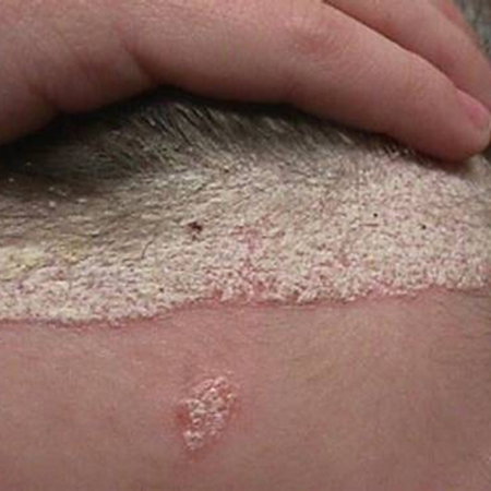 In patients without a diagnosis of psoriasis, skin inspection of the scalp, intergluteal fold, perineum, and periumbilical regions should be performed. The nails should be inspected for pitting or oncholysis.[Figure caption and citation for the preceding image starts]: Nail psoriasis: pitted nailsFrom the collection of Dr Tsu-Yi Chuang; used with permission [Citation ends].
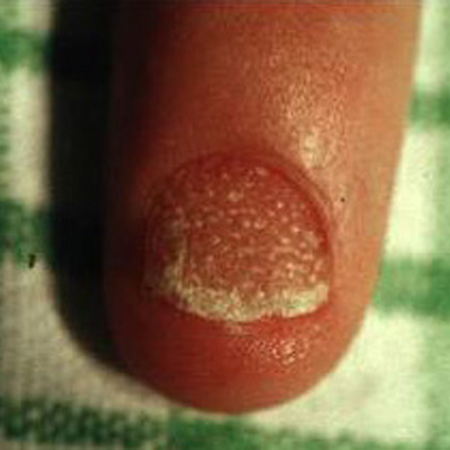
Musculoskeletal examination should include evaluation of all peripheral joints, including the interphalangeal joints of the fingers and toes, as well as the digits, for dactylitis. Swelling and tenderness of joints (synovitis) should be noted with examination of both the upper and lower extremities. Tenderness over the heel (either plantar fascia or Achilles tendon) and/or epicondyles of the elbow may indicate enthesitis.
Assessment of spine mobility is necessary to evaluate for spondylitis.[33] The occiput-to-wall or tragus-to-wall distance should be measured and cervical rotation assessed. A modified Schober test of the lumbar spine should also be performed. When performing this test, marks are made 5 cm below and 10 cm above a line at the 5th lumbar spinous process. The patient is asked to bend as far forward as possible. Normally, the distance between the upper and lower marks should increase by at least 5 cm. A distance less than this is consistent with diminished anteroposterior range of motion at the lumbar spine and suggests spinal stiffness.
Because of the increased risk of metabolic syndrome in psoriatic disease, blood pressure measurement and evaluation of obesity are an essential part of the physical examination.[34][35][36]
Assessment of disease extent
Although some patients may spontaneously go into remission, the majority of cases persist and potentially progress, leading to impaired function. A core assessment has been recommended for assessing the extent and severity of psoriatic disease at time of presentation and with each follow-up visit.[34][37][38]
Peripheral joint assessment; swollen and tender joint count.
Tender enthesitis count.
Skin assessment.
Documentation of patient's global assessment of disease activity and pain (achieved by using a visual analogue scale [VAS]).[39][Figure caption and citation for the preceding image starts]: Example of visual analogue pain scaleAdapted from Gould D, et al. Visual analogue scale (VAS). J Clin Nurs. 2001;10:706 [Citation ends].

Documentation of physician's global assessment of disease activity (also achieved using a VAS).
Standard assessment of physical function: Health Assessment Questionnaire (HAQ), Short Form 36 (SF-36), or PROMIS-Physical Function.[40][41][42]
Investigations
Patients with clinical evidence of joint inflammation should have plain film x-rays of the hands and feet since 40% of patients with PsA may have radiographic damage.[43] Erosion in the distal interphalangeal (DIP) joint and periarticular new-bone formation are suggestive of PsA. The classic changes of osteolysis leading to arthritis mutilans or a pencil-in-cup deformity are observed only in advanced disease. Soft-tissue swelling may be the only radiographic finding seen in early disease.
Initial laboratory investigations include rheumatoid factor (RF) and anti-cyclic citrullinated peptide (anti-CCP) antibody testing in patients with polyarthritis. Although RF can be positive in patients without rheumatoid arthritis (RA), a negative result may be of use in distinguishing PsA from RA (approximately 80% of RA patients will be positive for RF and anti-CCP antibodies).[44]
A high erythrocyte sedimentation rate (ESR) or C-reactive protein (CRP), although not specific for PsA (may be elevated in early RA in 40% of patients), is associated with polyarticular and progressive arthritis.[9][45][46] Because of the increased risk of metabolic syndrome in psoriatic disease, patients should have appropriate metabolic screening, including a lipid profile, fasting blood glucose, and a uric acid level. Synovial fluid aspiration and analysis may be necessary in patients with monoarthritis in order to exclude gout or infection.
Between 25% and 33% of patients with PsA have isolated spondylitis without sacroiliitis.[47][48] Asymmetrical sacroiliitis is characteristic of PsA and may be asymptomatic. X-ray findings in early disease are usually normal; however, radiographic changes in advanced disease can help to support the diagnosis. Therefore, plain film x-rays of the spine and pelvis are indicated in patients with symptoms or physical findings suggestive of hip, sacroiliac, or spine involvement.
Although not routinely indicated, an MRI scan of the sacroiliac joints may be of use in identifying those patients suspected of having early sacroiliitis despite the absence of radiographic changes.[4] Abnormal findings can occur in asymptomatic patients.
Joint ultrasound is currently a clinical research technique used to demonstrate the extent of entheseal involvement in psoriatic disease. Abnormalities demonstrated by ultrasound include tenosynovitis with abnormal fluid in the tendon sheath and disruption of tendon fibre architecture, erosions at the entheseal insertion site, and increased vascularity or Doppler signal.
Use of this content is subject to our disclaimer