Images and videos
Images
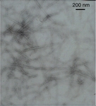
Amyloidosis
Electron micrograph demonstrating classical amyloid fibrils
Morie A. Gertz, MD; courtesy of Mayo Clinic
See this image in context in the following section/s:
Amyloidosis
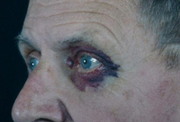
Bilateral periorbital ecchymosis (amyloid purpura) in a patient with AL amyloidosis
Williams MU, Murphy CE, Gore RS, et al. BMJ Case Rep 2018;11:e225923. doi:10.1136/bcr-2018- 225923
See this image in context in the following section/s:
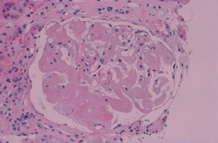
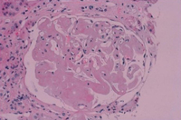
Amyloidosis
Renal biopsy demonstrating amyloid deposits as amorphous replacement of the glomerular architecture
Morie A. Gertz, MD; courtesy of Mayo Clinic
See this image in context in the following section/s:

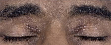
Amyloidosis
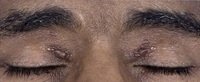
Classic periorbital purpura
Morie A. Gertz, MD; courtesy of Mayo Clinic
See this image in context in the following section/s:

Amyloidosis
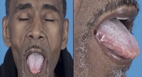
Macroglossia in a patient with AL amyloidosis
Williams MU, Murphy CE, Gore RS, et al. BMJ Case Rep 2018;11:e225923. doi:10.1136/bcr-2018- 225923
See this image in context in the following section/s:

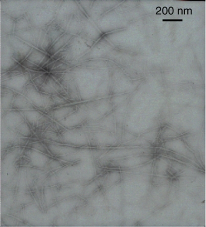
Amyloidosis
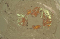
Congo red stain blood vessel in a bone marrow biopsy demonstrating green birefringence pathognomonic of amyloidosis
Morie A. Gertz, MD; courtesy of Mayo Clinic
See this image in context in the following section/s:
Use of this content is subject to our disclaimer