Approach
History
A complete history, with special attention to metabolic conditions, joint injuries and surgeries, kidney stones, diabetes mellitus, liver disease, and conditions associated with low magnesium levels, is recommended. The chronicity and pattern of the arthritis, and whether there are acute flares, should be documented.
Acute CPPD typically causes erythema, warmth, and swelling of the affected joint. Fever and constitutional symptoms may also be present. The knee is the most commonly affected joint, followed by the wrist.[14]
The most common form of calcium pyrophosphate (CPP) arthritis presents as a chronic degenerative arthritis that resembles osteoarthritis and may occur with or without inflammatory episodes. It typically affects joints not commonly affected by osteoarthritis, such as the wrists or shoulders.[14]
In addition to its presentation as an acute monoarticular or oligoarticular arthritis, CPP arthritis can present with a polyarticular, symmetric inflammatory arthritis similar to rheumatoid arthritis; or, less commonly, with the diffuse aching similar to that seen in polymyalgia rheumatica.
Queries about prolonged morning stiffness, diurnal variations in symptoms, and the effects of exercise on pain can be helpful in differentiating polyarticular CPP arthritis from rheumatoid arthritis or polymyalgia rheumatica.
Physical examination
Skin should be examined for abnormal pigmentation typical of haemochromatosis. A careful joint examination looking for bony deformities as well as any signs of acute inflammatory arthritis, such as warmth, redness, and synovial effusion, is recommended.
Synovial fluid analysis
This test is the cornerstone of diagnosis for CPP arthritis.[14] Arthrocentesis is therefore the key diagnostic manoeuvre. This safe and quick procedure can almost always be carried out at the bedside. Bacteraemia and active infection overlying the joint are the major contraindications to arthrocentesis. It can usually be performed safely in anticoagulated patients.
There are numerous sources describing proper approaches to aspirating joints. Generally, the joint should be entered at the joint space and major blood vessels avoided. With complex joints such as the mid-foot, or small joints such as the metatarsophalangeal joint, placing the needle in the area of maximal tenderness or fluctuance is often the most effective approach.

How to aspirate synovial fluid from the knee and administer intra-articular medication using a medial approach.
Fluid should be carefully examined under compensated polarised light microscopy for the presence of positively birefringent rhomboid-shaped crystals.[14] These crystals are sometimes rods or squares and can be weakly or even non-birefringent. A negative test does not rule out CPP arthritis, although the exclusive presence of urate crystals suggests gout as an alternative diagnosis. CPP crystals can be small, sparse, and are often weakly birefringent, and for these reasons can be easily overlooked.
If necessary, fluids can be refrigerated for several days before examination. Whether crystals are intracellular or extracellular has little influence on the diagnosis.[21] CPP crystals can be seen in joints from patients with acute CPP crystal arthritis well after their inflammation has resolved. [Figure caption and citation for the preceding image starts]: Images of intracellular calcium pyrophosphate crystals under compensated polarising light microscopyFrom the personal collection of Ann K. Rosenthal, MD [Citation ends].
X-rays
Plain x-rays of the affected joint can be helpful diagnostically.[46] Cartilage calcification can be seen in small joints, but patients with suspected polyarticular CPPD should have x-rays of the knees, pelvis, and wrist. These films have the highest likelihood of revealing radiographical cartilage calcification.[47] Plain x-rays can also rule out fractures, lytic bone lesions, and changes consistent with erosive rheumatoid arthritis. [Figure caption and citation for the preceding image starts]: Knee x-ray with linear calcific deposits of cartilage calcificationFrom the personal collection of Ann K. Rosenthal, MD [Citation ends].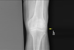 [Figure caption and citation for the preceding image starts]: Wrist radiograph from a patient with chronic calcium pyrophosphate arthritis showing severe degenerative changesFrom the personal collection of Ann K. Rosenthal, MD [Citation ends].
Blood tests
Blood tests to investigate the possibility of a co-existent metabolic condition should be performed in selected patients with premature or unusually widespread disease. Screening tests in these patients should include levels of serum calcium, magnesium, parathyroid hormone, alkaline phosphatase, ferritin, iron, serum transferrin saturation, and total iron-binding capacity.[14][15]
Erythrocyte sedimentation rate and C-reactive protein levels are often elevated during an acute attack of CPPD but have little diagnostic significance. Approximately 10% of patients with CPP arthritis have low levels of the rheumatoid factor (RF) autoantibody. Therefore, testing for RF is not typically useful in establishing the correct diagnosis.[1]
Specialist referrals
Patients should be referred to rheumatologists for refractory disease, if the practitioner is uncomfortable or unfamiliar with performing intra-articular injections or arthrocentesis, and if the diagnosis is unclear. Orthopaedic surgeons should be consulted for consideration of joint replacement surgery in people with incapacitating joint deformity or uncontrolled pain.
Other tests
When x-rays or joint aspiration are negative for CPPD but there is a strong clinical suspicion, ultrasound and CT scanning of affected joints may be confirmatory and can detect small calcific deposits sometimes missed by plain x-rays.[48][49] The double-contour sign on ultrasound identifies crystal-related arthropathies, but it cannot reliably distinguish between gout and CPPD in routine clinical practice. The diagnostic value is increased, however, when it is combined with hypervascularisation on ultrasound, and serum uric acid levels.[50]
Use of this content is subject to our disclaimer