Recommendations
Key Recommendations
The goal of treating wrist fractures is to optimise recovery of hand, wrist, and forearm function.[32][17]
Provide adequate analgesia and assess pain regularly.[31][33]
For adults, offer oral paracetamol (mild pain), oral paracetamol and codeine (moderate pain), or intravenous paracetamol with intravenous morphine (severe pain).[31]
For children under 16 years, offer oral ibuprofen and/or oral paracetamol (mild to moderate pain), or intranasal or intravenous opioids (moderate to severe pain).[31]
For unstable fractures and those requiring fixation, following closed reduction refer the patient immediately to a hand consultant, hand surgeon, or orthopaedic surgeon.
Casting is the definitive treatment for most forearm fractures in children.[33]
Elevate the affected limb.
For an open fracture, administer prophylactic intravenous antibiotics, ideally within 1 hour of injury.[35][47] Refer the patient to an orthopaedic or plastic surgeon. Do not irrigate open fractures.[34]
Refer the patient with suspected compartment syndrome immediately to orthopaedics.
Compartment syndrome is a surgical emergency and surgery should occur within 1 hour of the decision to operate.[48] See Compartment syndrome of extremities.
Assess and document any needs around safeguarding, falls risk, comorbidities, and the nature and classification of the fracture.[31]
Start rehabilitation of the hand at the earliest opportunity; patient education, reassurance, and pain control are essential during the initial visit.
Analgesia
Provide appropriate analgesia: fractures are typically associated with moderate to severe pain.
The type and dose of analgesia will vary with the amount of pain the patient is experiencing, the type and severity of injury, and other modifying factors (e.g., age, comorbidities, allergies). In the UK, the National Institute for Health and Care Excellence (NICE) recommends for the immediate management of pain in adults:[31]
Oral paracetamol for mild pain
Oral paracetamol and codeine for moderate pain
Intravenous paracetamol supplemented with intravenous morphine titrated to effect for severe pain.
In frail or older adults, use intravenous opioids with caution and do not offer non-steroidal anti-inflammatory drugs (NSAIDs). NSAIDs may be used as supplementary pain relief in other adults, who are not frail or elderly.[31]
In the UK, NICE recommends for the initial management of pain in children (under 16 years) with a suspected radial fracture:[31]
Oral ibuprofen, or oral paracetamol, or both for mild to moderate pain
Intranasal or intravenous opioids for moderate to severe pain (use intravenous opioids if intravenous access has been established).
These are often given at the time of the reduction for the sedative and analgesic effects.
Consider regional anaesthesia (haematoma block or peripheral nerve blockade), by healthcare professionals trained in the technique, when reducing a dorsally displaced radial fracture in adults in the emergency department.[31]
Do not give gas and air (nitrous oxide and oxygen) on its own in the emergency department for these fractures.[31]
Safeguarding
Assess for and document any needs around safeguarding, falls risk, comorbidities, and the nature and classification of the fracture.[31]
Closed fractures
If the patient is stable, apply a splint to the affected extremity to provide immobilisation and protection. Use either a full cast or a back slab, depending on the expertise of the person applying the splint and the preference of the patient.[17] In children, do not use a rigid cast for torus fractures of the distal radius.[31]
Refer undisplaced fractures and those that have been well reduced to the fracture clinic.
For unstable fractures and those requiring fixation, refer the patient immediately to a hand consultant, hand surgeon, or orthopaedic surgeon. If the fracture is displaced, urgent reduction may be performed in the emergency department by a clinician with the appropriate training and expertise.
Obtain post-reduction radiographs in the splint. If reduction is inadequate or unstable, an open reduction and fixation is likely to be necessary.
Once the fracture is immobilised, elevate the affected limb using a broad arm sling. Remove any rings as the hand will become swollen. Advise the patient to hold their wrist at the level of the heart when sitting. Encourage full-finger range of motion exercises while in the cast.
Practical tip
Be considerate when removing rings from the affected hand. Use a ring cutter when necessary; but there are techniques, such as using the elastic from an oxygen mask to compress the swollen tissue in the finger and assist with the removal of the ring, which may allow the ring to be safely removed without using a ring cutter. You might be able to place the ring onto the patient’s other hand for safekeeping.
Open fractures
In most situations, open fractures constitute a surgical emergency and operative treatment at the earliest possible opportunity is the preferred method of management. Immediately refer patients with open fractures and/or nerve compromise according to your local protocol. In the UK, this may be to the on-call orthopaedic team.
In the emergency department:[35][34]
Photograph the open fracture wound when it is first exposed for care
Keep the photographs in the patient’s records.
Follow your local protocol regarding taking, handling, and storing photographs and using them for clinical decision making.
Remove gross contamination
Do not irrigate open fractures
Prior to formal debridement, the wound should be handled only to remove gross contamination and to allow photography. ‘Mini-washouts’ outside the operating theatre environment are not indicated.[34]
Consider using a saline-soaked dressing covered with an occlusive layer
Administer prophylactic intravenous antibiotics, ideally within 1 hour of injury.[47]
Consider local antimicrobial resistance data when prescribing antibiotics. Follow your local protocol or take advice from microbiology.
Provide tetanus toxoid immunisation, if needed.[49]
More information: Tetanus toxoid immunisation
Consider providing a tetanus toxoid immunisation. This may involve a booster dose in a patient who has received an adequate priming course but whose last dose was more than 5 to 10 years ago. A patient who has not received an adequate priming course or is of uncertain immunisation status, or if there is heavy wound contamination, should receive intramuscular tetanus immunoglobulin and a reinforcing dose of vaccine.[49]
Realign and splint the limb.[34]
The fracture may be provisionally reduced in the emergency department by a clinician with the appropriate training and expertise.
This helps to reduce deformity and soft-tissue swelling, and may relieve any symptoms of nerve compression.
Consider referral for debridement, fixation, and soft tissue coverage of an open fracture by consultants in orthopaedic and plastic surgery.[35] Debridement should be performed:[35]
Immediately for highly contaminated open fractures
Within 12 hours of the injury for high-energy open fractures that are not highly contaminated
Within 24 hours of the injury for all other open fractures.
Open reduction and internal fixation is performed in the operating theatre. Thorough debridement and irrigation of the fracture and the open wound by the surgical team is required prior to fixation.[50] If the wound is grossly contaminated and there is a high concern for infection, internal fixation may be delayed or external fixation may be utilised as definitive treatment.[50] Use a temporary dressing that avoids wound desiccation and minimises the number of dressing changes after wound excision if immediate definitive soft tissue cover has not been performed.[35]
Most open fractures are high-energy injuries and may be accompanied by significant soft-tissue trauma. It is not uncommon for these patients to present with very swollen and tense forearms. Monitoring for median nerve function should be maintained throughout the postoperative period. Carpal tunnel decompression may be required if median nerve symptoms are present.
Open fractures are often obvious, but sometimes an apparently minor surface wound belies severe injury below. Therefore, any fracture associated with an overlying or nearby soft tissue injury, even an apparently innocuous minor wound, needs to be treated as an open fracture until shown otherwise.
Compartment syndrome
Evaluate the patient for forearm compartment syndrome. Refer the patient with suspected compartment syndrome immediately to orthopaedics.
Compartment syndrome is a surgical emergency and surgery should occur within 1 hour of the decision to operate.[36] See Compartment syndrome of extremities.
If there are obvious signs and symptoms of compartment syndrome, a clinical diagnosis is established and surgical fasciotomy is performed.[51]
Haemorrhage
Control frank haemorrhage with direct pressure or a tourniquet. Do not use blind clamping of bleeding.[37] Your local protocol should include combined review and decision-making in person by consultant surgeons skilled in vascular repair and skeletal trauma. The ischaemic limb should be revascularised within 4 hours from injury.[37]
For patients with severe acute haemorrhage (e.g., in the context of major trauma), consider antifibrinolytics (e.g., tranexamic acid). These agents have been shown to increase survival.[52][53] Delay in administration reduces their benefit; in a meta-analysis of data from patients with traumatic bleeding or post-partum haemorrhage, delays in administration of tranexamic acid were associated with reduced survival (survival benefit decreasing by about 10% for every 15 minutes of treatment delay until 3 hours, after which there was no benefit).[54]
A pulseless, deformed limb should be re-aligned and splinted. Repeat and document the vascular examination.[37] Refer to orthopaedic and vascular surgeons if the circulation is compromised.
Adults
Definitive treatment of a non-displaced fracture of the distal radius is a below-elbow cast for 4 to 6 weeks. A removable wrist splint may also be used.[55]
Non-displaced fractures of the distal radius usually result from low-energy injuries and are largely comfortable once the wrist is immobilised.
In patients with a stable fracture of the distal radius, consider early mobilisation from a removable support if pain allows.[32][17]
The choice of immobilisation may vary from a cast applied by the surgeon or a cast technician to a custom-made splint from an occupational therapist.
When using a plaster cast, the wrist should be positioned at neutral with three-point moulding used to hold the fracture, rather than forced palmar flexion.[32][17]
The thumb is free and the cast terminates at the level of the distal palmar flexion crease. This allows free motion of the metacarpophalangeal joints, thus maintaining digital mobility as the fracture heals, and minimises post-traumatic stiffness.
Casts should be well fitting and well padded to avoid any pressure effects, and the patient must be alerted to the possibility of needing a cast change. Cast changes may be necessary if the cast gets loose as the initial post-traumatic swelling reduces.
Consider removing the cast and starting mobilisation 4 weeks after the injury.[32][17]
Alternatively, in patients unable to tolerate casts or unwilling to wear a cast, or in patients who have an incomplete fracture of the distal radius, a forearm-based splint holding the wrist at neutral may be used.[56]
Splints are custom-made by occupational therapists, and can be custom-moulded to the patient's anatomy. As swelling reduces, modification to fit the changing dimensions of the patient's limb may be necessary.
Advise patients with non-displaced fractures or incomplete crack fractures of the radius about the possibility of spontaneous rupture of the extensor pollicis longus (EPL) tendon. This is a rare complication, with an incidence of 5% or less, that tends to occur within the first 12 weeks after injury and is usually preceded by increasing pain over the dorsal aspect of the distal radius.[57] Not all EPL ruptures are symptomatic and not all necessarily need to be treated.
Practical tip
To examine the EPL tendon, place the patient's hand flat on a table and ask the patient to lift only the thumb off the surface. With a rupture, the patient will be unable to raise the thumb in line with the second metacarpal.
[Figure caption and citation for the preceding image starts]: Cast treatment of a distal radius fractureFrom the collection of Dr Chaitanya S. Mudgal [Citation ends].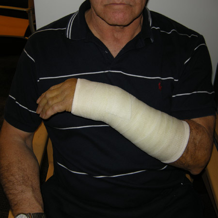
Children
Casting is the definitive treatment for most forearm fractures in children.[33] This is due to the greater capacity for remodelling following fracture union in children compared with adults.[33]
Do not use a rigid cast for torus fractures (also known as buckle fractures) of the distal radius.[33] A soft cast or bandaging may be used instead.[58]
One Cochrane review states that there is reassuring evidence of a full return to previous function with no serious adverse events, including refracture, for correctly diagnosed buckle fractures in children, whatever the treatment used, and that these findings are consistent with the move away from cast immobilisation for these injuries.[59]
Discharge children with a torus fracture after initial assessment; further review is usually not needed.[33] Remove the cast or bandaging as symptoms resolve, usually around 4 weeks later.
Adults
Definitive treatment of a simple displaced fracture of the distal radius consists of manipulation under anaesthetic and a below-elbow cast for 4 to 6 weeks. Complex fractures will require either closed reduction with K-wiring or, if unstable, open reduction and surgical fixation.
Restoration of anatomy is essential to maximise functional outcome.
Non-surgical management
Consider manipulation and a plaster cast or back slab in adults with dorsally displaced distal radius fractures.[31][17] The choice between a back slab and a full cast will depend on the expertise of the person applying the splint and the preference of the patient.[17]
In patients aged 65 years or older, consider non-operative treatment as the primary management for dorsally displaced distal radius fractures, unless there is significant deformity or neurological compromise.[32][17] Consider pre-injury function, medical comorbidities, and fracture characteristics when making a decision.[17]
Real-time image guidance may improve manipulations for distal radius fractures performed in the emergency department; however, there is no clear evidence in this area.[31]
When using a plaster cast, the wrist should be in neutral flexion with three-point moulding used to hold the fracture and not forced palmar flexion.[32] Consider removing the cast and starting mobilisation 4 weeks after injury.[32]
Consider regional anaesthesia (haematoma block, or peripheral nerve blockade), administered by healthcare professionals trained in the technique, when reducing dorsally displaced distal radius fractures (Colles’ fracture) in adults.[31][32] Reassure patients that digital numbness is to be expected.
Do not use gas and air (nitrous oxide and oxygen) alone.[31]
More complex fractures may require reproduction of the fracture deformity prior to reduction in order to mobilise the fracture site. Adequate reduction is verified by palpation for step-offs along the dorsal and radial surfaces. The fracture is then held in its reduced position in a well-moulded splint.
Surgical management
If the patient is unsuitable for prolonged casting or you observe inadequate reduction on imaging following manual reduction, consider surgical reduction and fixation. When surgery is required for a distal radius fracture, the UK National Institute for Health and Care Excellence (NICE) and the British Orthopaedic Association (BOA) recommend that it should be performed:[31][32]
Within 72 hours of the injury for an intra-articular fracture or for a re-displacement
Within 7 days of the injury for an extra-articular fracture.
Surgical fixation may involve closed reduction and casting, closed reduction and K-wire fixation or, if this is not possible, open reduction and internal fixation.[31][32] There is insufficient evidence from randomised controlled trials to determine the role of percutaneous pinning versus cast immobilisation alone.[60] Offer manipulation and cast if satisfactory reduction can be maintained in the plaster. Surgical fixation with K-wires has not been demonstrated to improve patients’ wrist function at 12 months compared with a cast.[61]
Offer K-wire fixation if no fracture of the articular surface of the radial carpal joint is detected, or if displacement of the radial carpal joint can be reduced by closed manipulation.[31]
Volar locking plate fixation was found to result in better fracture alignment than closed reduction and cast immobilisation; however, there were no clinically important differences between treatments with respect to patient-reported pain and function at 12 months post-treatment.[62]
Following volar plate fixation, patients can be safely treated with a soft dressing.[63] Some surgeons prefer a cast for pain management. However, this period should not be longer than 3 weeks.[64] Following K-wire fixation, a gauze is placed over the K-wires and a back slab applied. Wires are generally removed 4 to 6 weeks following surgery.
Following manual or surgical fracture reduction, if median nerve dysfunction develops or persists, a carpal tunnel release procedure should be performed urgently.
[Figure caption and citation for the preceding image starts]: Plate fixation after open reduction with a volarly placed plate and screwsFrom the collection of Dr Chaitanya S. Mudgal [Citation ends].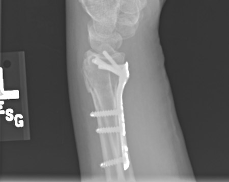
Assess the patient for falls risk and bone health, and refer as appropriate for any follow-up needed.[32] Explain to the patient what to expect about recovery and returning to normal activities, such as work, education, or driving.[32]
Children
In children, early, definitive manipulation and casting without admission is the standard of care:[33]
Manipulation of a child’s forearm fracture should be performed by competent orthopaedic practitioners.
Consider the analgesia necessary for this procedure (intranasal or intravenous opioids for moderate to severe pain).[31]
Manipulation of a child’s forearm fracture should be followed by orthogonal x-rays.
Assess the neurovascular status of the limb prior to discharge.
Provide oral analgesia to take home, along with information leaflets including information on any red flag symptoms, such as the cast being too tight (causing pain and swelling, which could create a compartment syndrome), or nerve symptoms such as pins and needles or loss of motor function.
A documented review of the case and images by a consultant orthopaedic surgeon should occur within 48 hours of injury.
For a child with a dorsally displaced distal radius fracture who has undergone manipulation, consider a below-elbow plaster cast or K-wire fixation if the fracture is completely displaced.[31]
Explain to the patient what to expect about recovery and returning to normal activities, such as education.[32]
Isolated non-displaced scaphoid fractures can be treated non-operatively in most patients, with high union rates and good clinical outcomes.[65][66]
Place the patient in a forearm-based cast without incorporating the thumb.[67] There is no universal consensus on the duration of casting, but usually the cast is maintained for a total of 6 to 8 weeks or until the fracture is healed.
Patients with proximal pole fractures or fracture displacement, or those who are unwilling to accept the protracted duration of casting, are considered candidates for percutaneous screw fixation or for open reduction and internal fixation of the scaphoid.[68][69]
For adult patients with scaphoid waist fractures displaced by 2 mm or less, consider an initial cast immobilisation. Any suspected non-unions can be confirmed and offered surgery.[70]
Non-displaced
Stable non-displaced fractures of the scaphoid and radius can in most instances be treated by non-operative means.[65][66]
Place the patient in a forearm-based cast without incorporating the thumb.[67] There is no universal consensus on the duration of casting, but usually the cast is maintained for a total of 6 to 8 weeks or until the fracture is healed.
Whether the patient is being treated in a cast or a splint or is awaiting surgical fixation, it is critical that they start rehabilitation of the hand at the earliest opportunity.
Patients with signs of carpal instability on radiography, proximal pole fractures, or fracture displacement, or those who are unwilling to accept the protracted duration of casting, are considered candidates for percutaneous screw fixation or for open reduction and internal fixation of the scaphoid.
Closed, displaced
It is essential to fix the scaphoid at the time of the radius fracture fixation. The primary surgical option is open reduction and fixation.[68] However, displaced fractures may also be treated with plate fixation.[71][72][73] Monitoring for median nerve function should be maintained throughout the postoperative period.
Provide the patient or carer with information on managing pain and oedema and recognising the signs and symptoms of common complications.[17][33] Refer patients presenting with excessive pain, oedema, loss of motion, or delayed functional recovery to physiotherapy or occupational therapy.[17]
Appropriate pain management is important, especially during rehabilitation; however, specific treatment varies widely depending on the patient, clinical presentation, method of treatment, and local treatment protocols.
Encourage the patient to use the injured limb while the wrist is immobilised for light functional activities, including self-care and tasks such as typing.[17] This is to help control oedema in the hand and to prevent stiffness in the metacarpophalangeal (MCP) and proximal interphalangeal joints and frozen shoulder.[74][75]
Consider early mobilisation with a removable support once pain allows in patients with a stable fracture of the distal radius.[17]
Consider a bone mineral density work-up in the orthopaedic clinic if needed. This can improve osteoporosis assessment and treatment rates following fragility fractures of the distal part of the radius.[48][17]
Use of this content is subject to our disclaimer