Aetiology
Falls on the outstretched hand are the most common cause of wrist fracture.[17] In older patients this is most often the causative mechanism and is usually a fall from standing height. In younger patients, falls commonly result from sporting activities or fractures may result from high-speed motor vehicle accidents.[1][12][13][14][15]
A similar mechanism of injury is also responsible for the causation of scaphoid fractures. However, it is the degree of radial or ulnar deviation of the wrist at the time of impact in addition to the degree of wrist extension and weight of the patient that ultimately determines the fracture pattern, be it of the radius or the scaphoid.
Pathophysiology
The type of fracture and degree of comminution are the resultant of several factors or variables:
the nature of the fall
the bone quality
the age and weight of the patient
the energy involved
the position of the hand and wrist at the time of impact.
Various combinations of these variables lead to a variety of different fracture patterns.
A fall on the outstretched hand causes extension of the wrist. The volar cortex of the distal radius therefore fails in tension and as such exhibits a simple fracture line. As the wrist continues to deform, the dorsal cortex fails in compression, leading to comminution of the dorsal cortex, a feature very commonly seen in most distal radius fractures and even more so in fractures associated with osteopenia/osteoporosis.
As the deformation of the wrist continues, the ulnar styloid may also fracture due to the attachment of the triangular fibrocartilage complex to the ulnar styloid. In most cases, the foveal insertion will still be intact, and consequently not all ulnar styloid fractures will lead to an unstable distal radioulnar joint. Therefore, most ulnar styloid fractures may be left untreated. In older women with osteoporosis, the distal ulna may fracture through the metaphysis. In such a situation the fracture pattern is that of a distal forearm fracture; please see our Long bone fracture topic for more information.
While it is easy for the patient to think of the distal radius as being part of the wrist and focus upon recovery of isolated wrist motion, in reality it is the alteration of forearm rotation after a distal radius fracture that can be the greatest source of symptoms: limitations of function, patient dissatisfaction, or both.
Classification
Association for the Study of Internal Fixation (AO/ASIF) classification[3]
A: Extra-articular fractures in which the fracture line does not extend into the radiocarpal or distal radioulnar joint (DRUJ).[Figure caption and citation for the preceding image starts]: Type A extra-articular fracture of the distal radius: lateral view [Citation ends].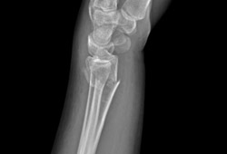
B: Partial intra-articular fracture in which the fracture line extends into the radiocarpal joint. Most commonly only a part of the articular surface is involved and the remaining part is still attached to the shaft.[Figure caption and citation for the preceding image starts]: Type B (simple) intra-articular fracture of the distal radius: lateral view [Citation ends].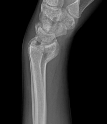
C: Intra-articular fracture in which the fracture line extends into the radiocarpal joint or the DRUJ.[Figure caption and citation for the preceding image starts]: Type C (complex) intra-articular fracture of the distal radius: lateral view [Citation ends].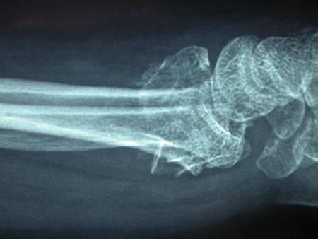
Each type is sub-classified into subgroups 1, 2, and 3, based on the severity of comminution or fracture location.
However, only the type of fracture (A, B, or C) provides substantial intra-observer reliability. The inclusion of subgroups into the classification has reduced the inter-observer and the intra-observer reliability to a level that is not reliable enough to use in clinical practice.[4][5]
Fernandez classification[6]
I: Bending of the metaphysis
II: Shearing fractures of the joint surface
III: Compression of the joint surface
IV: Avulsion or radiocarpal fracture dislocations
V: Combined fractures with high velocity injuries
This classification of the distal radius is based on the mechanism of injury.[7]
[Figure caption and citation for the preceding image starts]: Fernández classification (I: bending of the metaphysis; II: shearing fractures of the joint surface; III: compression of the joint surface; IV: avulsion or radiocarpal fracture dislocations; V: combined fractures with high velocity injuries)Shehovych A et al. Ann R Coll Surg Engl 2016 Nov;98(8):525-31; used with permission [Citation ends].
Frykman classification[8]
I: Extra-articular
II: As type I with fracture of the distal ulna
III: Radiocarpal joint involved
IV: As type III with fracture of the distal ulna
V: Distal radioulnar joint involved
VI: As type V with fracture of the distal ulna
VII: Radiocarpal and distal radioulnar joints both involved
VIII: As type VII with fracture of the distal ulna
This classification does not take into consideration the extent of fracture displacement, degree of comminution, or shortening of the radius, so its ability to guide treatment and predict prognosis is limited.[7]
[Figure caption and citation for the preceding image starts]: Frykman’s 1967 classification (I: extra-articular; II: as I with fracture of the distal ulna; III: radiocarpal joint involved; IV: as III with fracture of the distal ulna; V: distal radioulnar joint involved; VI: as V with fracture of the distal ulna; VII: radiocarpal and distal radioulnar joints both involved; VIII: as VII with fracture of the distal ulna)Shehovych A et al. Ann R Coll Surg Engl 2016 Nov;98(8):525-31; used with permission [Citation ends].
Use of this content is subject to our disclaimer