Images and videos
Images


Wrist fractures
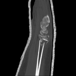
CT scans of the wrist provide excellent detail to assess fracture geometry, articular involvement, as well as degree of comminution
See this image in context in the following section/s:
Wrist fractures
Type C (complex) intra-articular fracture of the distal radius: lateral view
See this image in context in the following section/s:

Wrist fractures
Labelled version of previous figure. (a) 1=scaphoid; 2=lunate; 3=triquetral; 4-- =pisiform; 5a=body of hamate; 5b=hook of hamate; 6=capitate; 7=trapezoid; 8=trapezium; 1st→5th=respective metacarpal bases; r=radial styloid; u=ulnar styloid; (b) 1--=scaphoid; 2--=lunate; 6--=capitate; dr--=distal radius (note the distal ulna projected through this); t=dorsal tubercle of the radius; (c) as for a, except 5=hamate. *You can use the mnemonic “so long to pinky, here comes the thumb.” -- Object labelled within the dashed line
BMJ 2014;349:g5758; used with permission
See this image in context in the following section/s:
Wrist fractures
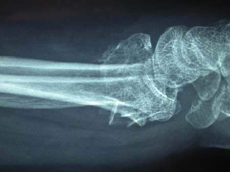
Type B (simple) intra-articular fracture of the distal radius: lateral view
See this image in context in the following section/s:

Wrist fractures
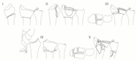
Fernández classification (I: bending of the metaphysis; II: shearing fractures of the joint surface; III: compression of the joint surface; IV: avulsion or radiocarpal fracture dislocations; V: combined fractures with high velocity injuries)
Shehovych A et al. Ann R Coll Surg Engl 2016 Nov;98(8):525-31; used with permission
See this image in context in the following section/s:

Wrist fractures
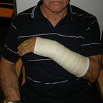
Cast treatment of a distal radius fracture
From the collection of Dr Chaitanya S. Mudgal
See this image in context in the following section/s:

Wrist fractures
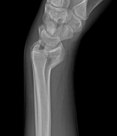
Type A extra-articular fracture of the distal radius: lateral view
See this image in context in the following section/s:
Wrist fractures
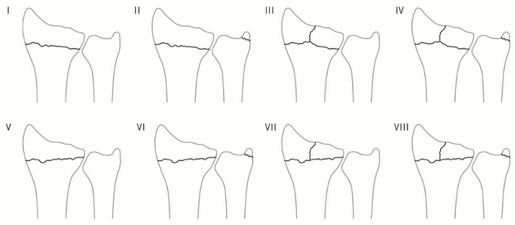
Frykman’s 1967 classification (I: extra-articular; II: as I with fracture of the distal ulna; III: radiocarpal joint involved; IV: as III with fracture of the distal ulna; V: distal radioulnar joint involved; VI: as V with fracture of the distal ulna; VII: radiocarpal and distal radioulnar joints both involved; VIII: as VII with fracture of the distal ulna)
Shehovych A et al. Ann R Coll Surg Engl 2016 Nov;98(8):525-31; used with permission
See this image in context in the following section/s:
Wrist fractures
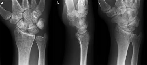
(a) Posteroanterior view, (b) lateral view, and (c) posteroanterior oblique view of a normal right wrist
BMJ 2014;349:g5758; used with permission
See this image in context in the following section/s:
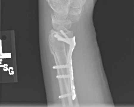
Wrist fractures
Plate fixation after open reduction with a volarly placed plate and screws
From the collection of Dr Chaitanya S. Mudgal
See this image in context in the following section/s:

Wrist fractures
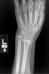
Posteroanterior x-ray showing malunion of the distal radius with significant shortening of the radius and relative lengthening of the ulna
See this image in context in the following section/s:
Use of this content is subject to our disclaimer