Images and videos
Images


Diabetes-related foot disease
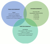
Diabetic foot problems can be related to the presence of a wound, ischaemia, or infection (WIfI). Which of these parameters is dominant can vary, and a flexible long-term management approach is needed. The Venn diagram shows intersecting rings of dominance for these three parameters, with gradings listed for each. The shaded areas represent combinations of these parameters of dominance
From the collection of Dr David G. Armstrong and Dr Joseph L. Mills Sr; used with permission
See this image in context in the following section/s:
Diabetes-related foot disease
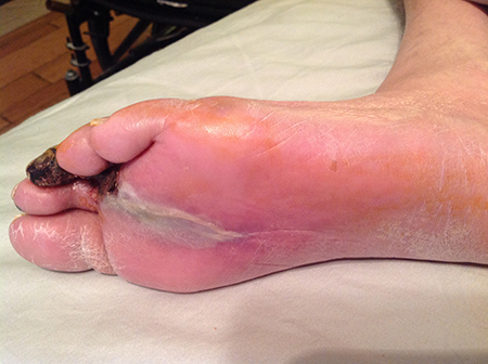
A foot infection originating from a gangrenous third toe. Note the erythema and fluctuance in the midfoot. An abscess cavity was found tracking under the longitudinal section of macerated skin
From the collection of Dr Neal R. Barshes; used with permission
See this image in context in the following section/s:
Diabetes-related foot disease
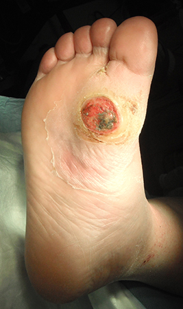
Uninfected foot ulcer overlying the plantar aspect of the first metatarsophalangeal joint. Note the hyperkeratotic skin (callus) surrounding the wound edge
From the collection of Dr Neal R. Barshes; used with permission
See this image in context in the following section/s:
Diabetes-related foot disease
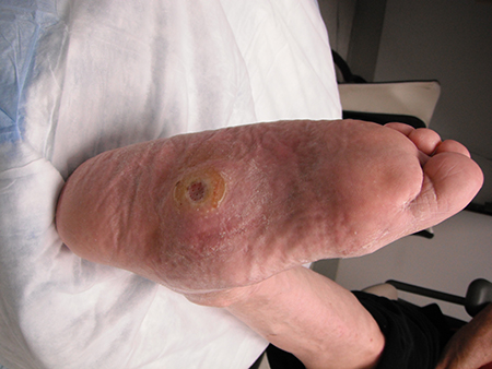
Midfoot ulcer in a patient with Charcot arthropathy (midfoot collapse)
From the collection of Dr Neal R. Barshes; used with permission
See this image in context in the following section/s:
Use of this content is subject to our disclaimer