Images and videos
Images


Interatrial communications (atrial septal defects)
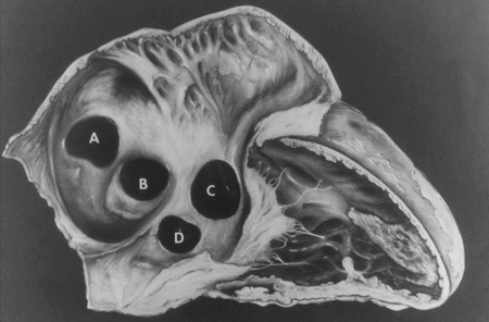
Subtypes of interatrial communications. A: sinus venosus defect; B: ostium secundum defect; C: ostium primum defect; D: unroofed coronary sinus defect
Mayo Clinic Foundation
See this image in context in the following section/s:

Interatrial communications (atrial septal defects)
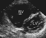
Parasternal short-axis echocardiographic image demonstrating right ventricular enlargement in a patient with an interatrial communication. LV: left ventricle; RV: right ventricle
Image courtesy of Patrick W. O'Leary, MD
See this image in context in the following section/s:
Interatrial communications (atrial septal defects)
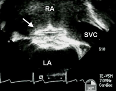
Trans-oesophageal echocardiographic image of an interatrial communication occluder device (arrow). LA: left atrium; RA: right atrium; SVC: superior vena cava
Image courtesy of Patrick W. O'Leary, MD
See this image in context in the following section/s:
Interatrial communications (atrial septal defects)
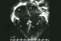
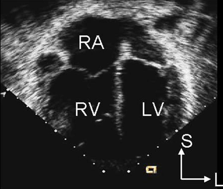
Apical 4-chamber echocardiographic image demonstrating right ventricular enlargement in a patient with an interatrial communication. L: lateral; LV: left ventricle; RA: right atrium; RV: right ventricle; S: superior
Image courtesy of Patrick W. O'Leary, MD
See this image in context in the following section/s:
Interatrial communications (atrial septal defects)
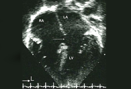
Apical 4-chamber echocardiographic image of an ostium primum defect (arrows). LA: left atrium; LV: left ventricle; RA: right atrium
Image courtesy of Patrick W. O'Leary, MD
See this image in context in the following section/s:
Use of this content is subject to our disclaimer