Images and videos
Images


Idiopathic intracranial hypertension
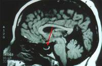
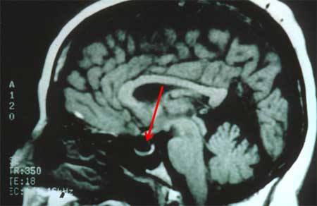
Magnetic resonance image (MRI) of empty sella on sagittal view
From the personal collection of Dr M. Wall; used with permission
See this image in context in the following section/s:
Idiopathic intracranial hypertension
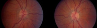
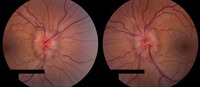
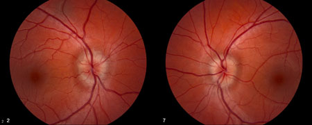
Bilateral optic atrophy
From the personal collection of Dr M. Wall; used with permission
See this image in context in the following section/s:
Idiopathic intracranial hypertension
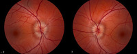
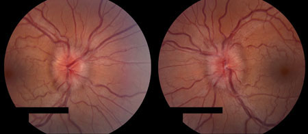
Frisén stage 2
From the personal collection of Dr M. Wall; used with permission
See this image in context in the following section/s:
Idiopathic intracranial hypertension
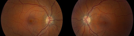
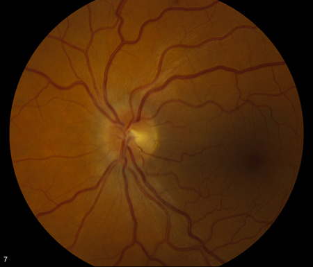
Frisén stage 1
From the personal collection of Dr M. Wall; used with permission
See this image in context in the following section/s:
Idiopathic intracranial hypertension
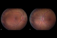
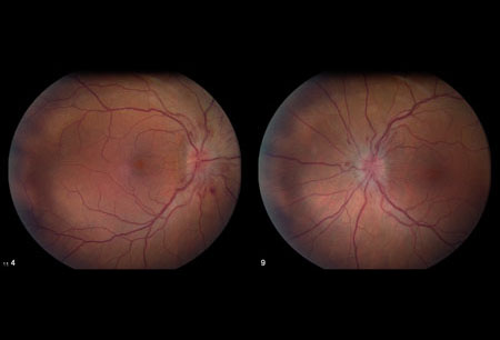
Bilateral disc oedema
From the personal collection of Dr M. Wall; used with permission
See this image in context in the following section/s:
Idiopathic intracranial hypertension
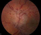
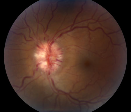
Frisén stage 3
From the personal collection of Dr M. Wall; used with permission
See this image in context in the following section/s:

Idiopathic intracranial hypertension
Frisén stage 5
From the personal collection of Dr M. Wall; used with permission
See this image in context in the following section/s:
Idiopathic intracranial hypertension
MRI of empty sella on sagittal view
From the personal collection of Dr M. Wall; used with permission
See this image in context in the following section/s:
Idiopathic intracranial hypertension
Frisén stage 4
From the personal collection of Dr M. Wall; used with permission
See this image in context in the following section/s:

Idiopathic intracranial hypertension
Bilateral disc swelling settled
From the personal collection of Dr M. Wall; used with permission
See this image in context in the following section/s:
Use of this content is subject to our disclaimer