Investigations
1st investigations to order
pure tone audiogram
Test
Performed in all patients to determine their hearing status.
Result
variable; commonly shows a conductive hearing loss; may also be normal or show a mixed conductive and sensorineural hearing loss in patients with cochlear damage or in those with a pre-existing hearing loss (e.g., congenital or presbycusis).
CT scan of the petrous temporal bones
Test
High-resolution CT scan of the petrous temporal bones is recommended as part of the initial work-up of patients with middle ear cholesteatoma.[25][26] It can provide confirmation of disease in patients with an atypical presentation, and can be used to assess the ear for mastoid pathology and complications such as cochlear, semicircular canal, or intracranial involvement.[27][Figure caption and citation for the preceding image starts]: Cholesteatoma, coronal CT scanFrom Dr Susan Douglas' personal teaching collection [Citation ends]. [Figure caption and citation for the preceding image starts]: Cholesteatoma, axial CT scanFrom Dr Susan Douglas' personal teaching collection [Citation ends].
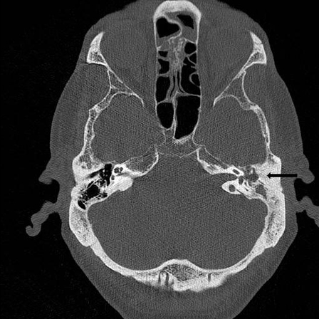 Intravenous contrast medium can be given if there is clinical suspicion of extra-osseous soft-tissue extension.[26]
Result
opacification of the middle ear or mastoid; with or without erosion of the scutum, ossicular chain, labyrinth, facial canal, tegmen, or bony capsule of the sigmoid sinus. CT with intravenous contrast may demonstrate the presence and extent of extra-osseous soft-tissue components.
Investigations to consider
fistula test
Test
Using tympanometry, pressure is applied to the tympanic membrane and eye movements are recorded with electronystagmography (ENG) electrodes.
Positive result is the production of nystagmus secondary to positive pressure applied to the tympanic membrane.
Result
variable; positive in presence of semicircular canal fistula; false-negatives may occur
MRI scan of the head and petrous temporal bones
Test
Should be ordered if there are suspected intracranial complications such as intracranial abscess, meningitis, or inner ear complications.[25]
MRI of the head with thin sections across the petrous temporal bone, pre- and post-intravenous contrast, helps to define the extent of the cholesteatoma prior to surgery.[26]
A non-echo-planar, fast-spin echo, or based diffusion-weighted MRI has been shown to be useful in the diagnosis of cholesteatoma. It is predominantly used in patients who have already had surgery in order to rule out recurrence or residual disease.[28][29][30]
Result
opacification of the middle ear and mastoid; may show evidence of intracranial complications such as a temporal lobe abscess, meningoencephalitis, intracranial extension, or sigmoid sinus thrombosis
bacterial culture
Test
Order if ear discharge is unresponsive to antimicrobial therapies.
Result
positive for micro-organisms
Use of this content is subject to our disclaimer