Images and videos
Images


Oesophageal cancer
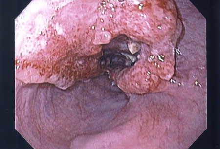
Endoscopic view of oesophageal cancer
Personal collection of Mark J. Krasna
See this image in context in the following section/s:

Oesophageal cancer
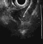
Endoscopic ultrasound-guided fine needle aspiration of lymph node
Personal collection of Mark J. Krasna
See this image in context in the following section/s:
Oesophageal cancer
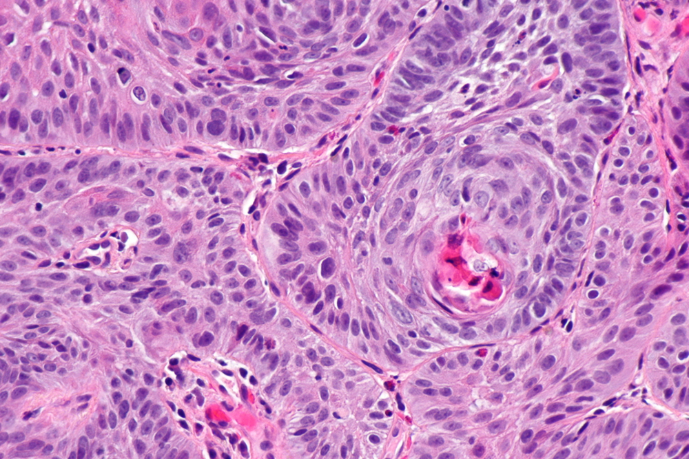
Moderated differentiated, keratinising oesophageal carcinoma
Wikimedia: Nephron https://creativecommons.org/licenses/by-sa/3.0/deed.en
See this image in context in the following section/s:
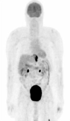
Oesophageal cancer
PET scan showing oesophageal cancer at the gastro-oesophageal junction. Note metastatic deposit in left femur
Personal collection of Mark J. Krasna
See this image in context in the following section/s:

Oesophageal cancer
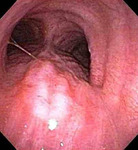
Tracheal invasion (T4) confirmed by bronchoscopy
Personal collection of Mark J. Krasna
See this image in context in the following section/s:
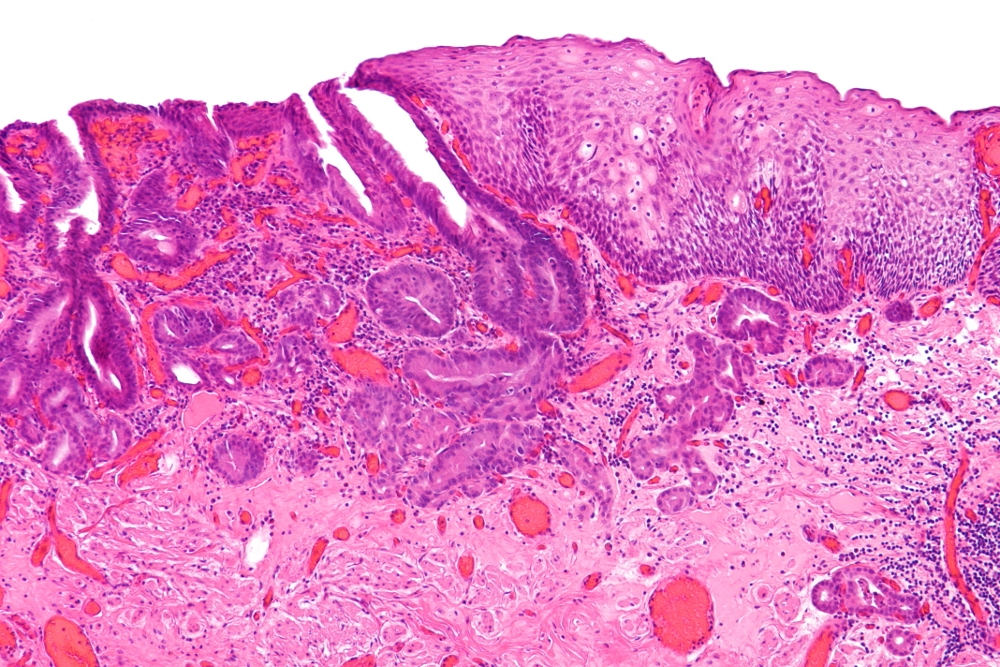
Oesophageal cancer
Adenocarcinoma (left of image) demonstrating glandular appearance with numerous mitotic cells and variable nuclear size and shape. Normal squamous epithelium is visible on the right of the image
Wikimedia: Nephron https://creativecommons.org/licenses/by-sa/3.0/deed.en
See this image in context in the following section/s:

Oesophageal cancer
CT scan showing T3 tumour at level of inferior pulmonary vein
Personal collection of Mark J. Krasna
See this image in context in the following section/s:
Use of this content is subject to our disclaimer