Images and videos
Images
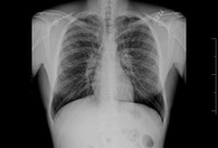
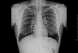
Bronchiectasis
Chest x-ray with dilated and thickened airways
From archives of Dr Sangeeta M. Bhorade; used with permission
See this image in context in the following section/s:

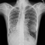
Bronchiectasis
Chest x-ray with lack of normal tapering producing a tram line
From archives of Dr Sangeeta M. Bhorade; used with permission
See this image in context in the following section/s:

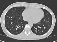
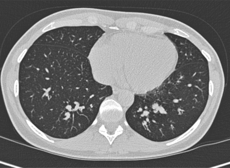
Bronchiectasis
Chest computed tomography scan with presence of signet ring on left
From archives of Dr Sangeeta M. Bhorade; used with permission
See this image in context in the following section/s:

Bronchiectasis
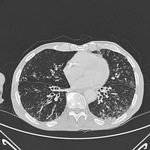
Severe cystic and varicose bronchiectasis in a 49-year-old man with idiopathic bronchiectasis and scoliosis
From Pamela J. McShane, MD; used with permission
See this image in context in the following section/s:
Bronchiectasis
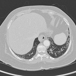
Mucus-impacted bronchi in a 36-year-old woman with allergic bronchopulmonary aspergillosis
From Pamela J. McShane, MD; used with permission
See this image in context in the following section/s:

Bronchiectasis
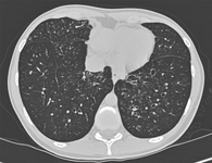
Signet ring signs in a 20-year-old woman with bronchiectasis
From Pamela J. McShane, MD; used with permission
See this image in context in the following section/s:

Bronchiectasis
Situs inversus (Kartagener syndrome) in a 19-year-old woman with focal bronchiectasis from primary ciliary dyskinesia
From Pamela J. McShane, MD; used with permission
See this image in context in the following section/s:

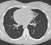
Bronchiectasis
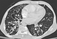
Chest computed tomography scan with dilated and thickened airways and peripheral tree-in-bud pattern
From archives of Dr Sangeeta M. Bhorade; used with permission
See this image in context in the following section/s:
Videos
 Expiratory wheeze
Auscultation sounds: Expiratory wheeze
 Early inspiratory crackles
Auscultation sounds: Early inspiratory crackles
Use of this content is subject to our disclaimer