Treatment algorithm
Please note that formulations/routes and doses may differ between drug names and brands, drug formularies, or locations. Treatment recommendations are specific to patient groups: see disclaimer
open partial fasciectomy + perioperative antibiotics
The most common procedure used in the surgical management of Dupuytren contracture, as it is associated with a postoperative recurrence rate of 15%.[49]
Through a transverse palmar incision overlying the distal palmar crease, the fascia forming pathologic cords is excised in a proximal-to-distal direction. [Figure caption and citation for the preceding image starts]: Preoperative view of the ring finger of a patient with a flexion contracture with surgical indications, showing the incision marking, demonstrating a transverse incision overlying the distal palmar crease, and oblique Brunner incisions coursing from it proximally and distallyFrom the collection of Dr C.M. Rodner; used with permission [Citation ends].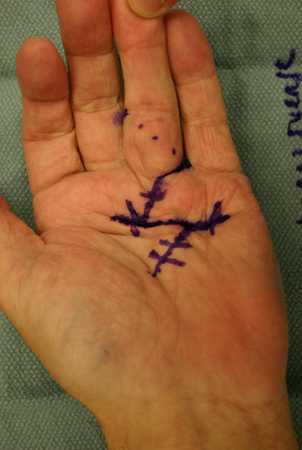 The choice of digital incision is surgeon-dependent and is most often between a Brunner and a longitudinal incision (often combined with multiple Z-plasties).
In the palm, the neurovascular structures deep to the involved fascia are identified and retracted. [Figure caption and citation for the preceding image starts]: Intraoperative view of the ring finger of a patient with a flexion contracture, with the radial digital neurovascular bundle identified and isolated coursing volar over the Dupuytren cord, which is being held up by forceps as it is excised in a proximal-to-distal directionFrom the collection of Dr C.M. Rodner; used with permission [Citation ends].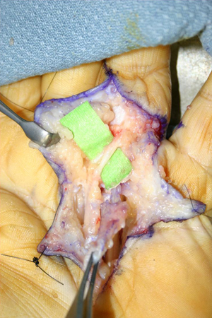 The diseased fascia is excised and elevated in a proximal-to-distal direction. As the Dupuytren cord moves into the finger, care is taken to identify the radial and ulnar digital arteries and nerves, particularly in PIP joint contractures. The joints are inspected for persistent contractures, and residual contracture of the PIP joint is addressed with a release of the volar plate followed by release of the collateral ligaments if necessary.
When possible, direct primary closure of the palmar skin is performed over a Penrose drain to prevent hematoma, as this method of closure allows for early motion and good skin sensibility, avoiding the meticulous wound care required with an open wound. [Figure caption and citation for the preceding image starts]: Postoperative view of the ring finger of a patient with a flexion contracture, showing the closed wound over a Penrose drain, which is used to minimize subsequent hematoma formationFrom the collection of Dr C.M. Rodner; used with permission [Citation ends]. If the palmar defect is too large for primary closure, skin grafting or the McCash "open-palm" technique are used. The patient is followed up within a few days to pull out the drain and assess the wound.
All patients with contractures should receive hand therapy postprocedure. After an open partial fasciectomy, flexion exercises begin once the wound has stabilized.
Postoperative infection is countered with the use of perioperative antibiotics and careful soft tissue handling. Cefazolin is the agent of choice. Clindamycin can be used as an alternative in patients with penicillin allergy, to provide gram-positive cover.
Primary options
cefazolin: 1g intravenously administered 30 minutes to 1 hour prior to the start of surgery
Secondary options
clindamycin: 600 mg intravenously administered 30 minutes to 1 hour prior to the start of surgery
postoperative splinting
Treatment recommended for SOME patients in selected patient group
After an open partial fasciectomy, the fingers can be splinted in full extension.
By the fifth postoperative day, patients are sent to the hand therapist for a forearm-based digital extension splint that is worn full time between therapy visits. Flexion exercises begin once the wound has stabilized. Regaining digital flexion often proves more difficult than maintaining extension after fasciectomy, due to the postoperative extension splinting required. [Figure caption and citation for the preceding image starts]: One-month postoperative view of the ring finger of a patient with a flexion contracture, demonstrating full active digital extensionFrom the collection of Dr C.M. Rodner; used with permission [Citation ends].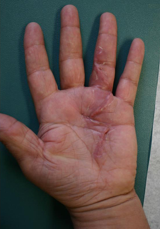 [Figure caption and citation for the preceding image starts]: One-month postoperative view of the ring finger of a patient with a flexion contracture, demonstrating active digital flexionFrom the collection of Dr C.M. Rodner; used with permission [Citation ends].
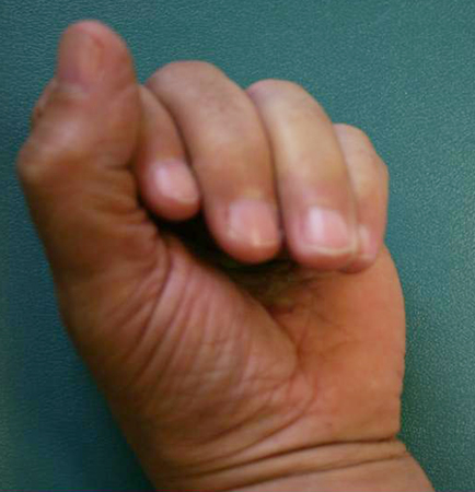 By the third postoperative week the splint is weaned, to be worn at night only, and nighttime extension splinting can continue for as long as 6 months.
Some surgeons have abandoned postoperative splinting, favoring earlier mobilization in order to minimize difficulties with flexion. There is also evidence to suggest that splinting (including nighttime extension splinting) after surgery provides no additional benefit to standard hand therapy in maintaining finger extension, except perhaps for cases in which extension loss occurs postoperatively, whereby nighttime extension splinting may provide some benefit.[53][54] Postoperative splinting may not be justified in all patients.
Choose a patient group to see our recommendations
Please note that formulations/routes and doses may differ between drug names and brands, drug formularies, or locations. Treatment recommendations are specific to patient groups. See disclaimer
Use of this content is subject to our disclaimer