Approach
The diagnosis of Dupuytren contracture is clinical and based on a thorough physical exam of the hands supported by an indicative clinical history.
Clinical history
Dupuytren contracture is typically seen in men of northern European descent aged >40 years. As it is believed to show autosomal dominant inheritance with variable penetrance, there may be a family history of the disease.[1] The medical history may be positive for diabetes mellitus or epilepsy, and the social history may reveal that the patient is a smoker or a heavy drinker.
Patients describe difficulties with face washing, combing their hair, and putting their hands in their pockets or fitting them into gloves.
Physical exam
A detailed physical exam of the affected hand(s) reveals a number of characteristic findings, depending on the disease progression. Bilateral hand involvement is common, with one hand usually more severely affected than the other, although the handedness of the patient is not a predictor of severity.
A firm and thickened palmar nodule over the metacarpal head at the level of the distal palmar crease, proximal to the metacarpophalangeal (MCP) joint, is often the earliest sign. This nodule may be associated with a band in the palmar aponeurosis. After nodule formation, palmar skin changes occur, with skin thickening, tethering, puckering, or pitting, as well as subcutaneous fat fibrosis.
The formation of pretendinous cords usually occurs as isolated nodules coalesce, although both nodules and cords can be present simultaneously. The most commonly affected digit is the ring finger, followed by the small finger, thumb, middle finger, and index finger. As pretendinous cords in the palm progress, they may travel across the MCP joint and, over time, produce MCP joint flexion contractures, leading to limited extension of the affected finger. [Figure caption and citation for the preceding image starts]: Preoperative view of a small finger flexion contracture with surgical indicationsFrom the collection of Dr C.M. Rodner; used with permission [Citation ends].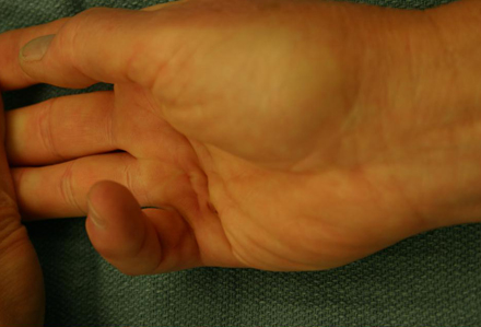 [Figure caption and citation for the preceding image starts]: Preoperative view of a small finger flexion contracture with surgical indicationsFrom the collection of Dr C.M. Rodner; used with permission [Citation ends].
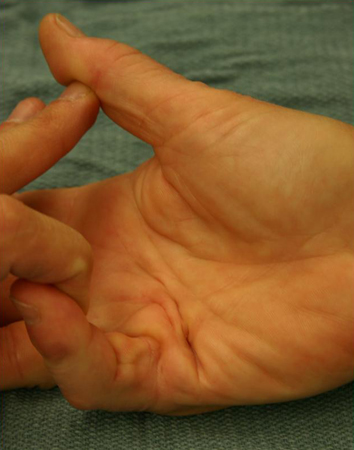 The degree of contracture is dependent on the severity of the disease. Digital cords that cross the proximal interphalangeal (PIP) joint may cause PIP joint contractures.
Exam of the dorsal aspect of the PIP joints may reveal areas of subcutaneous fibrosis, known as Garrod nodes or knuckle pads, which are indicative of systemic fascial disease and predictive of bilateral involvement. Garrod nodes are found in about one half of patients.
The term Dupuytren diathesis refers to patients with severe disease. These patients are usually younger, with very rapid disease progression involving both hands, and are more likely to have systemic fascial disease including Ledderhose disease affecting the plantar surface of the feet, and, in men, Peyronie disease, which affects the penis.[19]
The Hueston table-top test aids diagnosis and involves the patient attempting to lay the palm of the hand flat on a table surface. The test is positive if the patient is unable to flatten the hand on the table.
Investigations
As the diagnosis of Dupuytren contracture is predominantly clinical, ultrasound of the hand has limited usefulness in the diagnosis of the disease. It shows a mass lying between the flexor tendon below and the skin above. MRI and radiographs are not indicated in the diagnosis of the disease.
Use of this content is subject to our disclaimer