Images and videos
Images


Lipoma
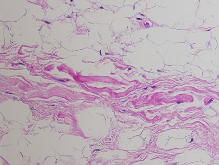
Spindle cell lipoma. Mature adipose tissue with intervening strands of dense fibrosis with spindle cell areas and characteristic ropey collagen bundles. Hematoxylin and eosin, 200x magnification
From the collection of Dr Kimberly Moore Dalal and Dr Steven D. DeMartini; used with permission
See this image in context in the following section/s:

Lipoma
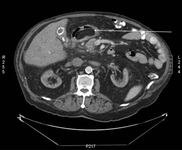
Gastric submucosal lipoma, CT scan. Submucosal antral mass with fatty density throughout.
From the collection of Dr Kimberly Moore Dalal and Dr Steven D. DeMartini; used with permission
See this image in context in the following section/s:
Lipoma
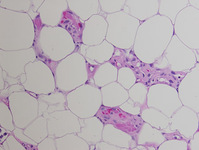
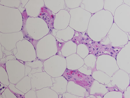
Angiolipoma. Mature adipose tissue with foci of endothelial proliferation containing microvascular thrombi. Hematoxylin and eosin, 200x magnification
From the collection of Dr Kimberly Moore Dalal and Dr Steven D. DeMartini; used with permission
See this image in context in the following section/s:

Lipoma
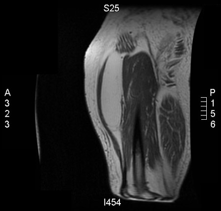
Intramuscular lipoma, right thigh. MRI, axial, T1-weighted image. Lipomatous mass in the anterior aspect of the right thigh
From the collection of Dr Kimberly Moore Dalal and Dr Steven D. DeMartini; used with permission
See this image in context in the following section/s:
Lipoma
Intramuscular lipoma, right thigh. MRI, coronal, T1-weighted image. Lipomatous mass in the anterior aspect of the right thigh
From the collection of Dr Kimberly Moore Dalal and Dr Steven D. DeMartini; used with permission
See this image in context in the following section/s:

Lipoma
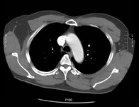
Intramuscular lipoma of subscapularis muscle, CT scan. Right axillary soft-tissue fatty mass with well-circumscribed margins
From the collection of Dr Kimberly Moore Dalal and Dr Steven D. DeMartini; used with permission
See this image in context in the following section/s:
Lipoma
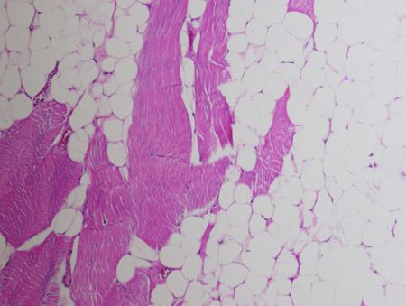
Intramuscular lipoma. Mature adipose tissue insinuating between skeletal muscle bundles. Hematoxylin and eosin, 200x magnification
From the collection of Dr Kimberly Moore Dalal and Dr Steven D. DeMartini; used with permission
See this image in context in the following section/s:
Lipoma
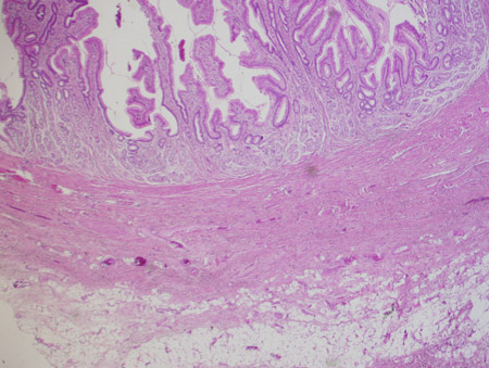
Gastric submucosal lipoma. A nodule of mature adipose tissue is present subjacent to gastric mucosa. Hematoxylin and eosin, 20x magnification
From the collection of Dr Kimberly Moore Dalal and Dr Steven D. DeMartini; used with permission
See this image in context in the following section/s:
Lipoma
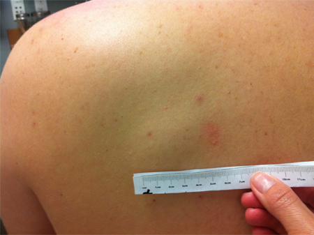
Subcutaneous lipoma on the trunk
From the collection of Dr Kimberly Moore Dalal and Dr Steven D. DeMartini; used with permission
See this image in context in the following section/s:

Lipoma
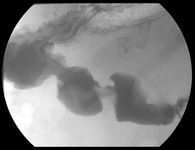
Gastric submucosal lipoma, upper GI contrast study. Filling defect in the distal antrum and pyloric channel suggesting antral mass prolapsing into pyloric channel
From the collection of Dr Kimberly Moore Dalal and Dr Steven D. DeMartini; used with permission
See this image in context in the following section/s:
Videos
 Intradermal injection animation demonstration
Demonstration of injection techniques used to administer local anesthetic, for allergy skin testing, and for tuberculin skin testing.
Use of this content is subject to our disclaimer