Etiology
The etiology for most lipomas is idiopathic. However, they may also appear on a hereditary basis in patients with familial multiple lipomatosis or in patients with Gardner syndrome.[9][10][21] Studies have also shown a correlation between HMG 1-C gene mutation and lipoma development.[22] Madelung disease, which features benign symmetric lipomatosis of the head, neck, shoulders, and proximal upper extremities, is associated with men with heavy alcohol consumption.[23] Dercum disease, also known as adiposis dolorosa, occurs in middle-aged women and is characterized by painful lipomas on the trunk, shoulders, arms, and legs; its etiology is unknown.[14] Other syndromes that may manifest lipomas include Bannayan-Riley-Ruvalcaba syndrome, Proteus syndrome, and multiple endocrine neoplasia 1.[24][25][26] Although trauma has been postulated as a potential inciting agent, it is unclear whether it is a true causal factor.[27][28]
Pathophysiology
Lipomas are slow-growing, benign, mesenchymal tumors that form well-circumscribed, lobulated lesions composed of adipocytes. They are demarcated from surrounding fat by a thin, fibrous capsule. Subcutaneous lesions are most common and are usually superficial, round, mobile, and soft, and feel similar to subcutaneous fat.
Gastrointestinal lipomas are uncommon and occur as submucosal lesions, most commonly in the stomach, small intestine, and colon.[7] They may present with intestinal obstruction or bleeding.[29] Rarely, lipomas can also occur in locations such as the adrenal glands, parotid glands, parapharyngeal space, breast, mediastinum, pleura, major airway, heart, superior vena cava, brain, and intraspinal areas.[8][Figure caption and citation for the preceding image starts]: Subcutaneous lipoma on the trunkFrom the collection of Dr Kimberly Moore Dalal and Dr Steven D. DeMartini; used with permission [Citation ends]. [Figure caption and citation for the preceding image starts]: Gastric submucosal lipoma, CT scan. Submucosal antral mass with fatty density throughout.From the collection of Dr Kimberly Moore Dalal and Dr Steven D. DeMartini; used with permission [Citation ends].
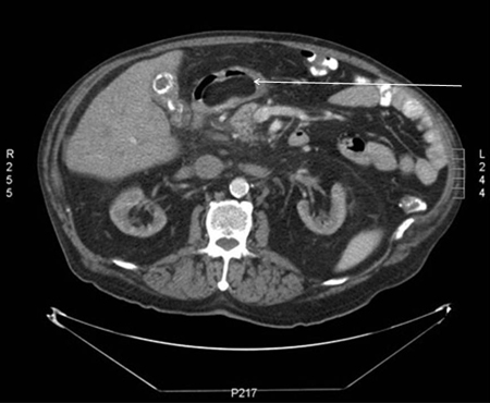 [Figure caption and citation for the preceding image starts]: Gastric submucosal lipoma, upper GI contrast study. Filling defect in the distal antrum and pyloric channel suggesting antral mass prolapsing into pyloric channelFrom the collection of Dr Kimberly Moore Dalal and Dr Steven D. DeMartini; used with permission [Citation ends].
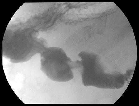
Different types of lipoma have specific histologic features.
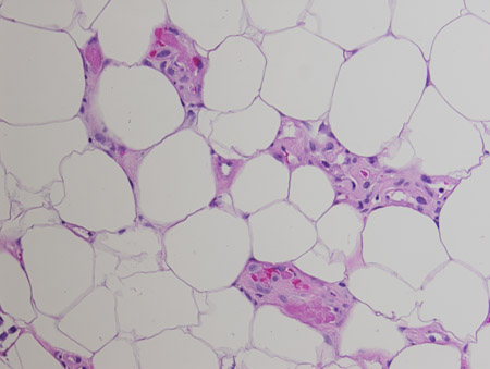
Angiolipomas are composed of adipocytes with interspersed clusters of capillaries containing fibrin thrombi. [Figure caption and citation for the preceding image starts]: Angiolipoma. Mature adipose tissue with foci of endothelial proliferation containing microvascular thrombi. Hematoxylin and eosin, 200x magnificationFrom the collection of Dr Kimberly Moore Dalal and Dr Steven D. DeMartini; used with permission [Citation ends].
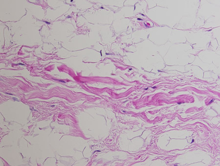
Spindle cell lipomas are composed of collagen-forming spindle cells that have replaced mature fat.[17][18][Figure caption and citation for the preceding image starts]: Spindle cell lipoma. Mature adipose tissue with intervening strands of dense fibrosis with spindle cell areas and characteristic ropey collagen bundles. Hematoxylin and eosin, 200x magnificationFrom the collection of Dr Kimberly Moore Dalal and Dr Steven D. DeMartini; used with permission [Citation ends].
Intramuscular lipomas are usually poorly circumscribed and infiltrative. With lesions in this anatomic position, it is important to exclude an atypical lipomatous tumor or well-differentiated liposarcoma as these are more common than a true lipoma in this position.[16][19] In order to confirm a diagnosis of atypical lipomatous tumor or well-differentiated liposarcoma, the excised specimen can be tested for MDM2 or CPM genes.[20][30] A diagnosis of atypical lipomatous tumor or well-differentiated liposarcoma should also be considered in patients with retroperitoneal lesions.[20]
Hibernomas resemble the glandular brown fat found in hibernating animals.[4][16] They have a greater tendency to bleed during excision and recur if not fully excised.
Classification
Clinical classification
The different types of lipoma are:
Superficial subcutaneous[Figure caption and citation for the preceding image starts]: Subcutaneous lipoma on the trunkFrom the collection of Dr Kimberly Moore Dalal and Dr Steven D. DeMartini; used with permission [Citation ends].
Intramuscular
Spindle cell: mature fat replaced by collagen-forming spindle cells
Angiolipoma: adipocytes interspersed with capillaries containing fibrin thrombi[2]
Lipoblastoma: variant found exclusively in infancy and early childhood[3]
Hibernoma: tumors consisting of glandular brown fat.[4]
Lipomas most commonly develop between 40 and 60 years of age, but congenital lipomas have been reported.[5][6]
Use of this content is subject to our disclaimer