Images and videos
Images


Acute cervical spine trauma
CT reconstruction demonstrating undisplaced odontoid fracture
From the personal collection of Michael G. Fehlings
See this image in context in the following section/s:
Acute cervical spine trauma
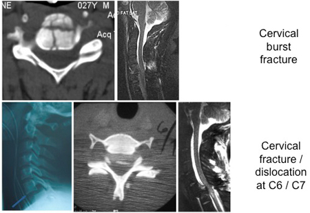
Common fracture patterns with severe cervical spine trauma. Top row: cervical burst fracture at C5 level; left: axial CT image showing a fracture of C5 vertebral body; right: mid-sagittal T2-weighted MRI showing retropulsion of the body of C5 with spinal cord compression, T2-weighted signal changes within the spinal cord and T2-weighted signal changes within the posterior ligamentous complex indicating disruption of these ligaments. Bottom row: fracture dislocation C6-C7 level. From left to right: lateral x-ray, axial CT through C6/C7 facet level, and T2-weighted mid-sagittal MRI demonstrating spinal cord compression and T2-weighted signal change within the spinal cord
From the personal collection of Michael G. Fehlings
See this image in context in the following section/s:
Acute cervical spine trauma
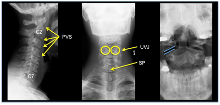
Normal cervical spine: lateral, AP, and open-mouth odontoid view
From the personal collection of Michael G. Fehlings
See this image in context in the following section/s:
Use of this content is subject to our disclaimer