Images and videos
Images
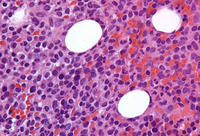


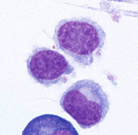
Hairy cell leukemia
Cytospin prepared from bone marrow aspirate illustrates the typical cell cytology, with oval- to bean-shaped nuclei and moderate amounts of cytoplasm with irregular cytoplasmic borders (Wright Giemsa 100×oil)
From the collection of Lynn Moscinski, MD
See this image in context in the following section/s:
Hairy cell leukemia
Sections of core biopsy demonstrate lymphocytes with obvious cytoplasm within the marrow interstitium, associated with dilation of marrow sinuses and red blood cell collections (H&E 50×oil)
From the collection of Lynn Moscinski, MD
See this image in context in the following section/s:
Use of this content is subject to our disclaimer