In the acute or subacute presentation, an initial assessment for the severity of the clinical condition is the first step, using the Airway, Breathing, Circulation, Disability, Exposure (ABCDE) protocol.[12]Resuscitation Council (UK). Resuscitation guidelines: the ABCDE approach. 2021 [internet publication].
https://www.resus.org.uk/library/abcde-approach
[13]Soar J, Böttiger BW, Carli P, et al. European Resuscitation Council guidelines 2021: adult advanced life support. Resuscitation. 2021 Apr;161:115-51.
https://www.resuscitationjournal.com/article/S0300-9572(21)00063-0/fulltext
http://www.ncbi.nlm.nih.gov/pubmed/33773825?tool=bestpractice.com
See the Urgent considerations section for immediate management for life-threatening conditions.
Time course
Acute dyspnea appears suddenly or in a matter of minutes. It typically indicates acute and severe conditions that may be life-threatening. Examples of conditions causing sudden-onset dyspnea include acute pulmonary embolism, myocardial infarction, acute heart valve insufficiency, pneumothorax, anaphylaxis, foreign body aspiration, pulmonary edema, or cardiac tamponade.[72]Roy CL, Minor MA, Brookhart MA, et al. Does this patient with a pericardial effusion have cardiac tamponade? JAMA. 2007 Apr 25;297(16):1810-8.
http://www.ncbi.nlm.nih.gov/pubmed/17456823?tool=bestpractice.com
Subacute dyspnea develops over hours to days. Common causes include acute asthma, exacerbation of chronic obstructive pulmonary disease (COPD), or pulmonary edema. Less common causes include myocarditis, superior vena cava syndrome, acute eosinophilic pneumonia, or cardiac tamponade.[72]Roy CL, Minor MA, Brookhart MA, et al. Does this patient with a pericardial effusion have cardiac tamponade? JAMA. 2007 Apr 25;297(16):1810-8.
http://www.ncbi.nlm.nih.gov/pubmed/17456823?tool=bestpractice.com
[73]Wilson LD, Detterbeck FC, Yahalom J. Clinical practice: superior vena cava syndrome with malignant causes. N Engl J Med. 2007 May 3;356(18):1862-9.
http://www.ncbi.nlm.nih.gov/pubmed/17476012?tool=bestpractice.com
[74]Allen JN, Magro CM, King MA. The eosinophilic pneumonias. Semin Respir Crit Care Med. 2002 Apr;23(2):127-34.
http://www.ncbi.nlm.nih.gov/pubmed/16088605?tool=bestpractice.com
Chronic dyspnea develops over weeks to months. It is associated with chronic pathology, such as congestive heart failure, COPD, cardiomyopathy, idiopathic pulmonary fibrosis, pulmonary vascular disease, pulmonary hypertension, valvular heart disease, or anemia.[75]Straus SE, McAlister FA, Sackett DL, et al; CARE-COAD1 Group. The accuracy of patient history, wheezing, and laryngeal measurements in diagnosing obstructive airway disease: JAMA. 2000 Apr 12;283(14):1853-7.
http://jamanetwork.com/journals/jama/fullarticle/192576
http://www.ncbi.nlm.nih.gov/pubmed/10770147?tool=bestpractice.com
Less common causes of chronic dyspnea include muscular dystrophies, kyphoscoliosis, amyotrophic lateral sclerosis, pulmonary alveolar proteinosis, chronic eosinophilic pneumonia, uremia, or constrictive pericarditis.[74]Allen JN, Magro CM, King MA. The eosinophilic pneumonias. Semin Respir Crit Care Med. 2002 Apr;23(2):127-34.
http://www.ncbi.nlm.nih.gov/pubmed/16088605?tool=bestpractice.com
[76]Sivak ED, Shefner JM, Sexton J. Neuromuscular disease and hypoventilation. Curr Opin Pulm Med. 1999 Nov;5(6):355-62.
http://www.ncbi.nlm.nih.gov/pubmed/10570736?tool=bestpractice.com
[77]Ferris G, Servera-Pieras E, Vergara P, et al. Kyphoscoliosis ventilatory insufficiency: noninvasive management outcomes. Am J Phys Med Rehabil. 2000 Jan-Feb;79(1):24-9.
http://www.ncbi.nlm.nih.gov/pubmed/10678599?tool=bestpractice.com
[78]Frazier AA, Franks TJ, Cooke EO, et al. From the archives of the AFIP: pulmonary alveolar proteinosis. Radiographics. 2008 May-Jun;28(3):883-99.
http://pubs.rsna.org/doi/full/10.1148/rg.283075219
http://www.ncbi.nlm.nih.gov/pubmed/18480490?tool=bestpractice.com
[79]Cimino JJ, Kogan AD. Constrictive pericarditis after cardiac surgery: report of three cases and review of the literature. Am Heart J. 1989 Dec;118(6):1292-301.
http://www.ncbi.nlm.nih.gov/pubmed/2686382?tool=bestpractice.com
An acute/subacute worsening of dyspnea in a patient with a known cardiovascular, pulmonary, or neuromuscular condition may represent a deterioration of the underlying condition or the appearance of a new problem.
Recurrent dyspnea may indicate paroxysmal tachycardias or intermittent complete heart block.
Clinical history
The approach to the patient with dyspnea depends on the severity of symptoms. Although careful history taking is crucial in the assessment of patients with all degrees of severity of dyspnea, this tends to be much more focused and concise for people presenting acutely. Similarly, in the acute presentation, the physical exam and investigations focus on vital findings that may point to (or rule out) life-threatening pathology. The approach to the patient with chronic dyspnea allows time for a more comprehensive, stepwise assessment.
The following factors need to be considered when taking a clinical history, although the time spent on asking questions and the degree of detail elicited will depend on the severity of the patient’s condition and the need for immediate investigations and treatment:
Severity
Dyspnea is highly subjective, and, for a given level of functional impairment, severity varies widely. There is no universally agreed measure of dyspnea; several scales are available in both research and clinical practice.[80]Johnson MJ, Oxberry SG, Cleland JG, et al. Measurement of breathlessness in clinical trials in patients with chronic heart failure: the need for a standardized approach: a systematic review. Eur J Heart Fail. 2010 Feb;12(2):137-47.
http://www.ncbi.nlm.nih.gov/pubmed/20083623?tool=bestpractice.com
Although scales exist, their use in everyday practice is limited.
Severe dyspnea in the acute presentation is typically associated with other symptoms and is more likely to be life-threatening (e.g., acute asthma, tension pneumothorax, acute upper airway obstruction, massive pulmonary embolism, or myocardial infarction).
Mild dyspnea may be a sole symptom and may indicate a benign etiology. It may be caused by stable COPD, deconditioning, noncritical airway obstruction, or normal aging.
In the more chronic presentation, measurement of severity using three domains is suggested: sensory-perceptual (measuring what breathing feels like); affecting distress (measuring how distressing breathing feels); and symptom impact (measuring how dyspnea affects functioning or quality of life).[1]Parshall MB, Schwartzstein RM, Adams L, et al. American Thoracic Society Committee on Dyspnea. An official American Thoracic Society statement: update on the mechanisms, assessment, and management of dyspnea. Am J Respir Crit Care Med. 2012 Feb 15;185(4):435-52.
http://www.atsjournals.org/doi/full/10.1164/rccm.201111-2042ST
http://www.ncbi.nlm.nih.gov/pubmed/22336677?tool=bestpractice.com
The Veterans Specific Activity Questionnaire (VSAQ) assesses the functional capability of the patient and estimates the aerobic capacity in meters.[81]Myers J, Bader D, Madhavan R, et al. Validation of a specific activity questionnaire to estimate exercise tolerance in patients referred for exercise testing. Am Heart J. 2001 Dec;142(6):1041-6.
http://www.ncbi.nlm.nih.gov/pubmed/11717610?tool=bestpractice.com
The degree of impairment can then be inferred. The approach and testing for a 25-year-old with trouble running an 8-minute mile is different from that of an 80-year-old with trouble climbing a 12-step staircase. The VSAQ establishes the baseline function and provides an objective measure for longitudinal assessment of progress or the lack thereof.
Other classification schemes include the New York Heart Association functional classification and the Medical Research Council dyspnea scale.[82]Goldman L, Hashimoto B, Cook EF, et al. Comparative reproducibility and validity of systems for assessing cardiovascular functional class: advantages of a new specific activity scale. Circulation. 1981 Dec;64(6):1227-34.
http://circ.ahajournals.org/content/64/6/1227
http://www.ncbi.nlm.nih.gov/pubmed/7296795?tool=bestpractice.com
[83]Mahler DA, Wells CK. Evaluation of clinical methods for rating dyspnea. Chest. 1988 Mar;93(3):580-6.
http://www.ncbi.nlm.nih.gov/pubmed/3342669?tool=bestpractice.com
Associated symptoms
Dyspnea often occurs with other symptoms, and their coexistence may help localize the origin of dyspnea to the involved organ system and help narrow the differential diagnosis.
Central chest pain is a common associated symptom in acute presentations and may suggest acute coronary syndrome, pulmonary embolism, pneumothorax, pneumomediastinum, or foreign body aspiration.[84]Caceres M, Ali SZ, Braud R, et al. Spontaneous pneumomediastinum: a comparative study and review of the literature. Ann Thorac Surg. 2008 Sep;86(3):962-6.
http://www.ncbi.nlm.nih.gov/pubmed/18721592?tool=bestpractice.com
A more chronic history of central chest pain on exertion, lasting less than 20 minutes, not rapidly increasing, and relieved by rest or nitroglycerin is more typical of stable angina.
Pleuritic chest pain may present acutely, subacutely, or chronically and may indicate pneumonia, pneumothorax, pulmonary embolism, a solitary fibrous tumor of the pleura, or pleuritis.[85]Rena O, Filosso PL, Papalia E, et al. Solitary fibrous tumour of the pleura: surgical treatment. Eur J Cardiothorac Surg. 2001 Feb;19(2):185-9.
http://www.ncbi.nlm.nih.gov/pubmed/11167110?tool=bestpractice.com
Pericardial constriction and effusions are characterized by typical pericardial pain that is referred to the scapular region, worsened by position and changes in intrathoracic pressure, and relieved by leaning forward.
Palpitations may be present in paroxysmal tachyarrhythmias, pulmonary embolism, valvular heart disease, or anxiety attacks.
Syncope may accompany dyspnea associated with tachyarrhythmias or pulmonary embolism.[49]Konstantinides SV, Meyer G, Becattini C, et al. 2019 ESC guidelines for the diagnosis and management of acute pulmonary embolism developed in collaboration with the European Respiratory Society (ERS): the Task Force for the diagnosis and management of acute pulmonary embolism of the European Society of Cardiology (ESC). Eur Respir J. 2019 Sep;543-603.
https://academic.oup.com/eurheartj/article/41/4/543/5556136
http://www.ncbi.nlm.nih.gov/pubmed/31504429?tool=bestpractice.com
Fever manifests with dyspnea in many infectious and inflammatory conditions, including pneumonia, bronchitis, laryngitis, viral syndromes (e.g., Hantavirus pulmonary syndrome and severe acute respiratory syndrome [SARS]), vasculitides, and sepsis.[86]Verity R, Prasad E, Grimsrud K, et al. Hantavirus pulmonary syndrome in northern Alberta, Canada: clinical and laboratory findings for 19 cases. Clin Infect Dis. 2000 Oct;31(4):942-6.
https://academic.oup.com/cid/article/31/4/942/376669/Hantavirus-Pulmonary-Syndrome-in-Northern-Alberta
http://www.ncbi.nlm.nih.gov/pubmed/11049774?tool=bestpractice.com
[87]Hui DS, Chan MC, Wu AK, et al. Severe acute respiratory syndrome (SARS): epidemiology and clinical features. Postgrad Med J. 2004 Jul;80(945):373-81.
http://pmj.bmj.com/content/80/945/373.long
http://www.ncbi.nlm.nih.gov/pubmed/15254300?tool=bestpractice.com
Dyspnea plus fever and cough may indicate community-acquired pneumonia or opportunistic infection in immunocompromised hosts. Postobstructive pneumonia is possible in patients with foreign body aspiration or a chest malignancy.
Fatigue, anorexia, nausea, myalgia, sore throat, expectoration of sputum, confusion, dizziness, gastrointestinal disturbance, anosmia, dysgeusia, rhinorrhea, and conjunctival congestion may occur with COVID-19. Patients who have recovered from COVID-19 may continue to experience dyspnea.[88]Fernández-de-Las-Peñas C, Palacios-Ceña D, Gómez-Mayordomo V, et al. Prevalence of post-COVID-19 symptoms in hospitalized and non-hospitalized COVID-19 survivors: a systematic review and meta-analysis. Eur J Intern Med. 2021 Oct;92:55-70.
https://www.ejinme.com/article/S0953-6205(21)00208-9/fulltext
http://www.ncbi.nlm.nih.gov/pubmed/34167876?tool=bestpractice.com
[89]Cabrera Martimbianco AL, Pacheco RL, Bagattini ÂM, et al. Frequency, signs and symptoms, and criteria adopted for long COVID-19: a systematic review. Int J Clin Pract. 2021 Oct;75(10):e14357.
https://www.ncbi.nlm.nih.gov/pmc/articles/PMC8236920
http://www.ncbi.nlm.nih.gov/pubmed/33977626?tool=bestpractice.com
Night sweats, malaise and weight loss may occur with tuberculosis infection.
Wheezing in the acute or subacute presentation may indicate acute asthma, acute exacerbation of COPD, pulmonary edema, bronchiolitis, or aspiration of a foreign body. Wheezing in the chronic presentation may indicate asthma, COPD, bronchiectasis, tracheobronchomalacia, or pulmonary or tracheobronchial tumors.
Cough may be present in acute bronchitis, acute infectious pneumonia, acute eosinophilic pneumonia, interstitial lung disease, COPD, asthma, bronchiectasis, tracheobronchomalacia, or chronic pneumonitis.[74]Allen JN, Magro CM, King MA. The eosinophilic pneumonias. Semin Respir Crit Care Med. 2002 Apr;23(2):127-34.
http://www.ncbi.nlm.nih.gov/pubmed/16088605?tool=bestpractice.com
Chronic sputum production may indicate COPD or bronchiectasis, while large amounts of clear secretions may be present in bronchoalveolar carcinoma.[90]Garfield DH, Cadranel JL, Wislez M, et al. The bronchioloalveolar carcinoma and peripheral adenocarcinoma spectrum of diseases. J Thorac Oncol. 2006 May;1(4):344-59.
http://www.ncbi.nlm.nih.gov/pubmed/17409882?tool=bestpractice.com
Change in the pitch of the voice may accompany dyspnea associated with pneumomediastinum, aortic aneurysm, retropharyngeal hematoma, lung cancer, or gastroesophageal reflux.[91]Mork T, Mutlu GM, Kuzniar TJ. Dysphonia and chest pain as presenting symptoms of pneumomediastinum. Tuberk Toraks. 2010;58(2):184-7.
http://www.ncbi.nlm.nih.gov/pubmed/20865573?tool=bestpractice.com
[92]Chen JW, Vela MF, Peterson KA, et al. AGA clinical practice update on the diagnosis and management of extraesophageal gastroesophageal reflux disease: expert review. Clin Gastroenterol Hepatol. 2023 Jun;21(6):1414-21.
https://www.cghjournal.org/article/S1542-3565(23)00143-X/fulltext
http://www.ncbi.nlm.nih.gov/pubmed/37061897?tool=bestpractice.com
Hemoptysis may accompany dyspnea in patients with COVID-19, acute bronchitis, exacerbation of bronchiectasis, chest malignancies, vasculitides, acute infectious pneumonia, cryptogenic organizing pneumonia, pulmonary embolism, cocaine toxicity, tuberculosis, or diffuse alveolar hemorrhage.[93]Hamilton W, Sharp D. Diagnosis of lung cancer in primary care: a structured review. Fam Pract. 2004 Dec;21(6):605-11.
https://academic.oup.com/fampra/article/21/6/605/508696/Diagnosis-of-lung-cancer-in-primary-care-a
http://www.ncbi.nlm.nih.gov/pubmed/15520035?tool=bestpractice.com
[94]Holguin F, Ramadan B, Gal AA, et al. Prognostic factors for hospital mortality and ICU admission in patients with ANCA-related pulmonary vasculitis. Am J Med Sci. 2008 Oct;336(4):321-6.
http://www.ncbi.nlm.nih.gov/pubmed/18854674?tool=bestpractice.com
[95]Oymak FS, Demirbas HM, Mavili E, et al. Bronchiolitis obliterans organizing pneumonia: clinical and roentgenological features in 26 cases. Respiration. 2005 May-Jun;72(3):254-62.
http://www.ncbi.nlm.nih.gov/pubmed/15942294?tool=bestpractice.com
[96]Mancano A, Marchiori E, Zanetti G, et al. Pulmonary complications of crack cocaine use: high-resolution computed tomography of the chest. J Bras Pneumol. 2008 May;34(5):323-7.
http://www.scielo.br/scielo.php?script=sci_arttext&pid=S1806-37132008000500012&lng=en&nrm=iso&tlng=en
http://www.ncbi.nlm.nih.gov/pubmed/18545829?tool=bestpractice.com
[97]Weber AM, Areerat P, Fischer JE, et al. Factors associated with diagnostic evaluation for tuberculosis among adults hospitalized for clinical pneumonia in Thailand. Infect Control Hosp Epidemiol. 2008 Jul;29(7):648-57.
http://www.ncbi.nlm.nih.gov/pubmed/18564918?tool=bestpractice.com
[98]Sachdeva A, Matuschak GM. Diffuse alveolar hemorrhage following alemtuzumab. Chest. 2008 Jun;133(6):1476-78.
http://www.ncbi.nlm.nih.gov/pubmed/18574290?tool=bestpractice.com
Dysphagia or odynophagia may be present in a dyspneic patient with granulomatous laryngitis, pneumomediastinum, foreign body aspiration, tetanus, and epiglottitis.[99]Silva L, Damrose E, Bairao F, et al. Infectious granulomatous laryngitis: a retrospective study of 24 cases. Eur Arch Otorhinolaryngol. 2008 Jun;265(6):675-80.
http://www.ncbi.nlm.nih.gov/pubmed/18060554?tool=bestpractice.com
[100]Bunch TJ, Thalji MK, Pellikka PA, et al. Respiratory failure in tetanus: case report and review of a 25-year experience. Chest. 2002 Oct;122(4):1488-92.
http://www.ncbi.nlm.nih.gov/pubmed/12377887?tool=bestpractice.com
[101]Wick F, Ballmer PE, Haller A. Acute epiglottis in adults. Swiss Med Wkly. 2002 Oct 12;132(37-38):541-7.
http://www.ncbi.nlm.nih.gov/pubmed/12557859?tool=bestpractice.com
In epiglottitis, dyspnea may be additionally accompanied by drooling. Vomiting and diarrhea may accompany dyspnea in thyrotoxicosis or botulism.[102]Varma JK, Katsitadze G, Moiscrafishvili M, et al. Signs and symptoms predictive of death in patients with foodborne botulism - Republic of Georgia, 1980-2002. Clin Infect Dis. 2004 Aug 1;39(3):357-62.
https://academic.oup.com/cid/article/39/3/357/352455/Signs-and-Symptoms-Predictive-of-Death-in-Patients
http://www.ncbi.nlm.nih.gov/pubmed/15307002?tool=bestpractice.com
[103]Guleria R, Goswami R, Shah P, et al. Dyspnoea, lung function & respiratory muscle pressures in patients with Graves' disease. Indian J Med Res. 1996 Nov;104:299-303.
http://www.ncbi.nlm.nih.gov/pubmed/8979522?tool=bestpractice.com
Heartburn may be present in gastroesophageal reflux with aspiration.
Muscle weakness or myalgias associated with dyspnea may indicate deconditioning, adverse effects of medications, muscular dystrophies, amyotrophic lateral sclerosis, acute polio or postpolio syndrome, Guillain-Barre syndrome, West Nile and other viral infections, leptospirosis, Cushing myopathy, or botulism.[76]Sivak ED, Shefner JM, Sexton J. Neuromuscular disease and hypoventilation. Curr Opin Pulm Med. 1999 Nov;5(6):355-62.
http://www.ncbi.nlm.nih.gov/pubmed/10570736?tool=bestpractice.com
[87]Hui DS, Chan MC, Wu AK, et al. Severe acute respiratory syndrome (SARS): epidemiology and clinical features. Postgrad Med J. 2004 Jul;80(945):373-81.
http://pmj.bmj.com/content/80/945/373.long
http://www.ncbi.nlm.nih.gov/pubmed/15254300?tool=bestpractice.com
[102]Varma JK, Katsitadze G, Moiscrafishvili M, et al. Signs and symptoms predictive of death in patients with foodborne botulism - Republic of Georgia, 1980-2002. Clin Infect Dis. 2004 Aug 1;39(3):357-62.
https://academic.oup.com/cid/article/39/3/357/352455/Signs-and-Symptoms-Predictive-of-Death-in-Patients
http://www.ncbi.nlm.nih.gov/pubmed/15307002?tool=bestpractice.com
[104]Boltan DD, Lachar W, Khetan A, et al. Fatal and widespread skeletal myopathy confirmed morphologically years after initiation of simvastatin therapy. Am J Cardiol. 2007 Apr 15;99(8):1171-6.
http://www.ncbi.nlm.nih.gov/pubmed/17437752?tool=bestpractice.com
[105]Benditt JO. Respiratory complications of amyotrophic lateral sclerosis. Semin Respir Crit Care Med. 2002 Jun;23(3):239-47.
http://www.ncbi.nlm.nih.gov/pubmed/16088616?tool=bestpractice.com
[106]Wijdicks EF, Henderson RD, McClelland RL. Emergency intubation for respiratory failure in Guillain-Barré syndrome. Arch Neurol. 2003 Jul;60(7):947-8.
http://jamanetwork.com/journals/jamaneurology/fullarticle/784408
http://www.ncbi.nlm.nih.gov/pubmed/12873850?tool=bestpractice.com
[107]Mutlu GM, Kuzniar T, Factor P. A 41-year-old man with altered mental status and acute flaccid paralysis. Chest. 2005 Jan;127(1):391-4.
http://www.ncbi.nlm.nih.gov/pubmed/15654004?tool=bestpractice.com
[108]Vieira SR, Brauner JS. Leptospirosis as a cause of acute respiratory failure: clinical features and outcome in 35 critical care patients. Braz J Infect Dis. 2002 Jun;6(3):135-9.
http://www.scielo.br/scielo.php?script=sci_arttext&pid=S1413-86702002000300006&lng=en&nrm=iso&tlng=en
http://www.ncbi.nlm.nih.gov/pubmed/12144750?tool=bestpractice.com
[109]Blanco C, Marazuela M, Flores J, et al. Severe respiratory failure secondary to Cushing's myopathy. J Endocrinol Invest. 2001 Sep;24(8):618-21.
http://www.ncbi.nlm.nih.gov/pubmed/11686545?tool=bestpractice.com
Visual disturbances may occur with dyspnea in myasthenia and tetanus, and headache may be present in carbon monoxide poisoning.[100]Bunch TJ, Thalji MK, Pellikka PA, et al. Respiratory failure in tetanus: case report and review of a 25-year experience. Chest. 2002 Oct;122(4):1488-92.
http://www.ncbi.nlm.nih.gov/pubmed/12377887?tool=bestpractice.com
[110]Hopkins LC. Clinical features of myasthenia gravis. Neurol Clin. 1994 May;12(2):243-61.
http://www.ncbi.nlm.nih.gov/pubmed/8041340?tool=bestpractice.com
[111]Abelsohn A, Sanborn MD, Jessiman BJ, et al. Identifying and managing adverse environmental health effects: 6. Carbon monoxide poisoning. CMAJ. 2002 Jun 25;166(13):1685-90.
http://www.cmaj.ca/content/166/13/1685.full
http://www.ncbi.nlm.nih.gov/pubmed/12126326?tool=bestpractice.com
Bone pain may be associated with acute chest syndrome due to sickle cell anemia or fat embolism associated with long-bone fractures.[112]Johnson CS. The acute chest syndrome. Hematol Oncol Clin North Am. 2005 Oct;19(5):857-79.
http://www.ncbi.nlm.nih.gov/pubmed/16214648?tool=bestpractice.com
Anxiety may be a reaction to dyspnea of any etiology but may also cause dyspnea in acute panic or anxiety attacks.[113]Periyakoil VS, Skultety K, Sheikh J. Panic, anxiety, and chronic dyspnea. J Palliat Med. 2005 Apr;8(2):453-9.
http://www.ncbi.nlm.nih.gov/pubmed/15890059?tool=bestpractice.com
Dyspnea associated with stress may indicate anxiety, hyperventilation, or takotsubo cardiomyopathy.[114]Sealove BA, Tiyyagura S, Fuster V. Takotsubo cardiomyopathy. J Gen Intern Med. 2008 Nov;23(11):1904-8.
http://www.ncbi.nlm.nih.gov/pubmed/18688681?tool=bestpractice.com
Abdominal pain, nausea, vomiting and diarrhea may occur in patients with e-cigarettes, or vaping product use associated lung injury (EVALI). Gastrointestinal symptoms may precede respiratory symptoms of dyspnea, cough and chest pain.[115]Kalininskiy A, Bach CT, Nacca NE, et al. E-cigarette, or vaping, product use associated lung injury (EVALI): case series and diagnostic approach. Lancet Respir Med. 2019 Dec;7(12):1017-26.
http://www.ncbi.nlm.nih.gov/pubmed/31711871?tool=bestpractice.com
[116]Cao DJ, Aldy K, Hsu S, et al. Review of Health Consequences of Electronic Cigarettes and the Outbreak of Electronic Cigarette, or Vaping, Product Use-Associated Lung Injury. J Med Toxicol. 2020 Jul;16(3):295-310.
https://www.doi.org/10.1007/s13181-020-00772-w
http://www.ncbi.nlm.nih.gov/pubmed/32301069?tool=bestpractice.com
[117]Evans ME, Twentyman E, Click ES, et al. Update: interim guidance for health care professionals evaluating and caring for patients with suspected e-cigarette, or vaping, product use-associated lung injury and for reducing the risk for rehospitalization and death following hospital discharge - United States, December 2019. MMWR Morb Mortal Wkly Rep. 2020 Jan 3;68(5152):1189-94.
https://www.ncbi.nlm.nih.gov/pmc/articles/PMC6943965
Positionality
Orthopnea is the presence of dyspnea while supine, with an improvement in the upright position. It is characteristically linked with congestive heart failure but may also be present in asthma, COPD, inflammatory and degenerative neurologic diseases, gastroesophageal reflux, pericardial effusion, or bilateral diaphragmatic paralysis.[76]Sivak ED, Shefner JM, Sexton J. Neuromuscular disease and hypoventilation. Curr Opin Pulm Med. 1999 Nov;5(6):355-62.
http://www.ncbi.nlm.nih.gov/pubmed/10570736?tool=bestpractice.com
[118]Greub G, Liaudet L, Wiesel P, et al. Respiratory complications of gastroesophageal reflux associated with paraesophageal hiatal hernia. J Clin Gastroenterol. 2003 Aug;37(2):129-31.
http://www.ncbi.nlm.nih.gov/pubmed/12869882?tool=bestpractice.com
[119]Torchio R, Gulotta C, Greco-Lucchina P, et al. Orthopnea and tidal expiratory flow limitation in chronic heart failure. Chest. 2006 Aug;130(2):472-9.
http://www.ncbi.nlm.nih.gov/pubmed/16899847?tool=bestpractice.com
[120]Celli BR, Rassulo J, Corral R. Ventilatory muscle dysfunction in patients with bilateral idiopathic diaphragmatic paralysis: reversal by intermittent external negative pressure ventilation. Am Rev Respir Dis. 1987 Nov;136(5):1276-8.
http://www.ncbi.nlm.nih.gov/pubmed/3674587?tool=bestpractice.com
Platypnea is the worsening of dyspnea on assuming an upright position, with alleviation while supine. It is typical of patent foramen ovale, abdominal muscle deficiency, or hepatopulmonary syndrome.[121]Kedia G, Tobis J, Lee MS. Patent foramen ovale: clinical manifestations and treatment. Rev Cardiovasc Med. 2008 Summer;9(3):168-73.
http://www.ncbi.nlm.nih.gov/pubmed/18953276?tool=bestpractice.com
[122]Lange PA, Stoller JK. The hepatopulmonary syndrome. Ann Intern Med.1995 Apr 1;122(7):521-9.
http://www.ncbi.nlm.nih.gov/pubmed/7872588?tool=bestpractice.com
Trepopnea is an infrequent finding where dyspnea is present only in the lateral decubitus position. It is associated with congestive heart failure, sinus of Valsalva aneurysms, or after a pneumonectomy.[123]Acosta J, Khan F, Chitkara R. Trepopnea resulting from large aneurysm of sinus of Valsalva and descending aorta. Heart Lung. 1982 Jul-Aug;11(4):342-4.
http://www.ncbi.nlm.nih.gov/pubmed/6919530?tool=bestpractice.com
[124]Alfaifi S, Lapinsky SE. Trepopnea due to interatrial shunt following lung resection. Chest. 1998 Jun;113(6):1726-7.
http://www.ncbi.nlm.nih.gov/pubmed/9631823?tool=bestpractice.com
Variable positional changes in dyspnea may also be seen in primary and metastatic cardiac tumors.[125]Tufekcioglu O, Yildiz A, Kacmaz F, et al. Trepopnea in a patient with cardiac tumor. Echocardiography. 2006 Feb;23(2):165-7.
http://www.ncbi.nlm.nih.gov/pubmed/16445740?tool=bestpractice.com
[126]Leung RS, Bowman ME, Parker JD, et al. Avoidance of the left lateral decubitus position during sleep in patients with heart failure: relationship to cardiac size and function. J Am Coll Cardiol. 2003 Jan 15;41(2):227-30.
http://www.ncbi.nlm.nih.gov/pubmed/12535814?tool=bestpractice.com
Pattern of dyspnea
Dyspnea that appears during the working week and resolves over periods off work may be related to occupational exposure and suggests occupational asthma.[127]Malo JL, Ghezzo H, L'Archeveque J, et al. Is the clinical history a satisfactory means of diagnosing occupational asthma? Am Rev Respir Dis. 1991 Mar;143(3):528-32.
http://www.ncbi.nlm.nih.gov/pubmed/2001062?tool=bestpractice.com
[128]Barber CM, Cullinan P, Feary J, et al. British Thoracic Society clinical statement on occupational asthma. Thorax. 2022 May;77(5):433-42.
https://thorax.bmj.com/content/77/5/433
Occupational exposure may also be implicated in cases of asbestos-related lung disease and hypersensitivity pneumonitis.[129]Markowitz SB, Morabia A, Lilis R, et al. Clinical predictors of mortality from asbestosis in the North American Insulator Cohort, 1981 to 1991. Am J Respir Crit Care Med. 1997 Jul;156(1):101-8.
http://www.atsjournals.org/doi/full/10.1164/ajrccm.156.1.9610108
http://www.ncbi.nlm.nih.gov/pubmed/9230732?tool=bestpractice.com
A history of occupational or leisure exposure to aerosolized solvents, fumes, organic dust, molds, and animals should be elicited and may be implicated in interstitial lung disease. Dyspnea developing in indoor hockey players may reflect nitrogen dioxide or carbon monoxide toxicity from faulty ice resurfacing equipment.[130]Karlson-Stiber C, Hojer J, Sjoholm A, et al. Nitrogen dioxide pneumonitis in ice hockey players. J Intern Med. 1996 May;239(5):451-6.
http://www.ncbi.nlm.nih.gov/pubmed/8642238?tool=bestpractice.com
Seasonal dyspnea or shortness of breath related to cold, pets, exercise, or nonspecific irritants may suggest asthma or reactive airway disease.[131]Lucero PF, Nicholson KL, Haislip GD, et al. Increased airway hyperreactivity with the M40 protective mask in exercise-induced bronchospasm. J Asthma. 2006 Dec;43(10):759-63.
http://www.ncbi.nlm.nih.gov/pubmed/17169828?tool=bestpractice.com
Dyspnea occurring from the day prior to menses, and up to three days after menses, may indicate a catamenial pneumothorax.[132]Gil Y, Tulandi T. Diagnosis and treatment of catamenial pneumothorax: a systematic review. J Minim Invasive Gynecol. 2020 Jan;27(1):48-53.
http://www.ncbi.nlm.nih.gov/pubmed/31401265?tool=bestpractice.com
Past medical history
It is important to consider the possibility of comorbidities.
Asthma may be an extraesophageal manifestation of gastroesophageal reflux disease, while COPD often coexists with other conditions, such as cardiovascular diseases, gastroesophageal reflux, and lung cancer, which can make the differential diagnosis difficult.[25]Global Initiative for Chronic Obstructive Lung Disease. Global strategy for prevention, diagnosis and management of COPD: 2025 report. 2024 [internet publication].
https://goldcopd.org/2025-gold-report
[92]Chen JW, Vela MF, Peterson KA, et al. AGA clinical practice update on the diagnosis and management of extraesophageal gastroesophageal reflux disease: expert review. Clin Gastroenterol Hepatol. 2023 Jun;21(6):1414-21.
https://www.cghjournal.org/article/S1542-3565(23)00143-X/fulltext
http://www.ncbi.nlm.nih.gov/pubmed/37061897?tool=bestpractice.com
Dyspnea may be associated with obesity or occur during a normal pregnancy. In pregnant patients dyspnea may indicate the presence of a previously undiagnosed medical condition, such as valvular heart disease, pulmonary hypertension, alpha-1 protease inhibitor (alpha-1 antitrypsin) deficiency, pulmonary embolism, spontaneous pneumothorax or pneumomediastinum, progression of a pulmonary arteriovenous malformation, or deterioration of myasthenia.[133]Anderson JA, Kennelly MM. Successful management of antenatal presentation of cor triatriatum. Eur J Obstet Gynecol Reprod Biol. 2008 Sep;140(1):137-8.
http://www.ncbi.nlm.nih.gov/pubmed/17977640?tool=bestpractice.com
[134]Bonnin M, Mercier FJ, Sitbon O, et al. Severe pulmonary hypertension during pregnancy: mode of delivery and anesthetic management of 15 consecutive cases. Anesthesiology. 2005 Jun;102(6):1133-7.
http://www.ncbi.nlm.nih.gov/pubmed/15915025?tool=bestpractice.com
[135]Giesler CF, Buehler JH, Depp R. Alpha1-antitrypsin deficiency: severe obstructive lung disease and pregnancy. Obstet Gynecol. 1977 Jan;49(1):31-4.
http://www.ncbi.nlm.nih.gov/pubmed/299781?tool=bestpractice.com
[136]Sellman JS, Holman RL. Thromboembolism during pregnancy: risks, challenges, and recommendations. Postgrad Med. 2000 Sep 15;108(4):71-2, 77-8, 81-4.
http://www.ncbi.nlm.nih.gov/pubmed/11021260?tool=bestpractice.com
[137]Tanase Y, Yamada T, Kawaryu Y, et al. A case of spontaneous pneumothorax during pregnancy and review of the literature. Kobe J Med Sci. 2007;53(5):251-5.
http://www.med.kobe-u.ac.jp/journal/contents/53/251.pdf
http://www.ncbi.nlm.nih.gov/pubmed/18204301?tool=bestpractice.com
[138]Esplin MS, Varner MW. Progression of pulmonary arteriovenous malformation during pregnancy: case report and review of the literature. Obstet Gynecol Surv. 1997 Apr;52(4):248-53.
http://www.ncbi.nlm.nih.gov/pubmed/9095491?tool=bestpractice.com
[139]Benshushan A, Rojansky N, Weinstein D. Myasthenia gravis and preeclampsia. Isr J Med Sci. 1994 Mar;30(3):229-33.
http://www.ncbi.nlm.nih.gov/pubmed/8181923?tool=bestpractice.com
[140]Strnad P, McElvaney NG, Lomas DA. Alpha(1)-Antitrypsin Deficiency. N Engl J Med. 2020 Apr 9;382(15):1443-55.
In a patient who is or recently was in labor, dyspnea may indicate pulmonary embolism, septic or toxic shock, amniotic fluid or trophoblastic embolism, pneumothorax, or pneumomediastinum.[50]Habek D, Habek JC. Nonhemorrhagic primary obstetric shock. Fetal Diagn Ther. 2008;23(2):140-5.
http://www.ncbi.nlm.nih.gov/pubmed/18046073?tool=bestpractice.com
[141]Zapardiel I, Delafuente-Valero J, Diaz-Miguel V, et al. Pneumomediastinum during the fourth stage of labor. Gynecol Obstet Invest. 2009;67(1):70-2.
http://www.ncbi.nlm.nih.gov/pubmed/18843189?tool=bestpractice.com
Dyspnea in the postoperative period may indicate pulmonary embolism, an acute coronary event, or pulmonary edema related to fluid resuscitation. Less frequently a pneumothorax or previously unrecognized muscular dystrophy may be implicated.[142]Souayah N, Tick Chong PS, Dreyer M, et al. Myotonic dystrophy type 1 presenting with ventilatory failure. J Clin Neuromuscul Dis. 2007 Sep;9(1):252-5.
http://www.ncbi.nlm.nih.gov/pubmed/17989589?tool=bestpractice.com
Specific surgical interventions may be followed by dyspnea due to fat embolism (liposuction, long-bone surgery), diaphragmatic paralysis (aortic valve surgery, lung surgery, and coronary artery bypass surgery), talc-induced acute lung injury (pleurodesis), or pulmonary vein stenosis (mitral valve surgery).[51]Wessman DE, Kim TT, Parrish JS. Acute respiratory distress following liposuction. Mil Med. 2007 Jun;172(6):666-8.
http://www.ncbi.nlm.nih.gov/pubmed/17615855?tool=bestpractice.com
[143]Kuzniar TJ, Blum MG, Kasibowska-Kuzniar K, et al. Predictors of acute lung injury and severe hypoxemia in patients undergoing operative talc pleurodesis. Ann Thorac Surg. 2006 Dec;82(6):1976-81.
http://www.ncbi.nlm.nih.gov/pubmed/17126094?tool=bestpractice.com
[144]Lange TJ, Luchner A, Endemann D, et al. A 46-year-old man with dyspnea and hemoptysis 3 years following mitral valve repair. Chest. 2007 Aug;132(2):704-7.
http://www.ncbi.nlm.nih.gov/pubmed/17699145?tool=bestpractice.com
A history of previous venothromboembolic disease, inadequate anticoagulation, immobilization, admission to the hospital, long-distance travel, vascular access, or leg injury may indicate pulmonary embolism as the cause of dyspnea.
The presence of a known autoimmune or rheumatologic disease predisposes the patient to dyspnea resulting from pulmonary embolism, pulmonary hypertension, interstitial lung disease, pleural effusion, or pulmonary hemorrhage.[145]Cha SI, Fessler MB, Cool CD, et al. Lymphoid interstitial pneumonia: clinical features, associations and prognosis. Eur Respir J. 2006 Aug;28(2):364-9.
http://erj.ersjournals.com/content/28/2/364.full
http://www.ncbi.nlm.nih.gov/pubmed/16571614?tool=bestpractice.com
Known malignancy may cause dyspnea through airway obstruction by the primary or metastatic tumor, malignant effusion, postobstructive pneumonia, pulmonary tumor embolism, lymphangitic spread into the lung, or pericardial or endocardial involvement.[146]Storck K, Crispens M, Brader K. Squamous cell carcinoma of the cervix presenting as lymphangitic carcinomatosis: a case report and review of the literature. Gynecol Oncol. 2004 Sep;94(3):825-8.
http://www.ncbi.nlm.nih.gov/pubmed/15350381?tool=bestpractice.com
[147]Moreno-Vega AL, Fuentes-Pradera J, Gordon-Santiago Mdel M, et al. Intraventricular metastases from rectal-sigmoid adenocarcinoma. Clin Transl Oncol. 2006 Apr;8(4):296-7.
http://www.ncbi.nlm.nih.gov/pubmed/16648108?tool=bestpractice.com
History of rheumatologic diseases, prior thromboembolic disease, uncorrected obstructive sleep apnea, and obesity may indicate pulmonary hypertension.
Recurrent pneumonia may indicate gastroesophageal reflux with aspiration, a retained foreign body, benign or malignant tumors, or a vascular ring.[148]Mukhopadhyay S, Katzenstein AL. Pulmonary disease due to aspiration of food and other particulate matter: a clinicopathologic study of 59 cases diagnosed on biopsy or resection specimens. Am J Surg Pathol. 2007 May;31(5):752-9.
http://www.ncbi.nlm.nih.gov/pubmed/17460460?tool=bestpractice.com
[149]Derkay CS, Wiatrak B. Recurrent respiratory papillomatosis: a review. Laryngoscope. 2008 Jul;118(7):1236-47.
http://www.ncbi.nlm.nih.gov/pubmed/18496162?tool=bestpractice.com
[150]Grathwohl KW, Afifi AY, Dillard TA, et al. Vascular rings of the thoracic aorta in adults. Am Surg. 1999 Nov;65(11):1077-83.
http://www.ncbi.nlm.nih.gov/pubmed/10551760?tool=bestpractice.com
Pleural effusions can accompany pneumonia, heart failure, pleural tuberculosis, malignancy, rheumatologic diseases, or mesothelioma.
A history of thoracic radiation for malignancy should be elicited. Dyspnea due to radiation pneumonitis typically appears 1 to 6 months after the radiation treatments.[151]Madani I, De Ruyck K, Goeminne H, et al. Predicting risk of radiation-induced lung injury. J Thorac Oncol. 2007 Sep;2(9):864-74.
http://www.ncbi.nlm.nih.gov/pubmed/17805067?tool=bestpractice.com
History of prior endotracheal intubation and prolonged mechanical ventilation may suggest subglottic scarring and stenosis.
Dyspnea in a person with history of COVID may be a symptom of long COVID.[152]Michelen M, Manoharan L, Elkheir N, et al. Characterising long COVID: a living systematic review. BMJ Glob Health. 2021 Sep;6(9):e005427.
https://www.ncbi.nlm.nih.gov/pmc/articles/PMC8478580
http://www.ncbi.nlm.nih.gov/pubmed/34580069?tool=bestpractice.com
Drug history
Medications may cause dyspnea or contribute to it through a variety of mechanisms, including several forms of pulmonary toxicity (interstitial disease, pulmonary edema, pulmonary hemorrhage, airways disease, pleural effusion, pulmonary vascular changes), induction of metabolic acidosis (nucleoside reverse-transcriptase inhibitors, topiramate), or induction of bradycardia and chronotropic insufficiency (digoxin, calcium-channel blockers, beta-blockers).
Pneumotox on line: patterns - interstitial/parenchymal lung disease
Opens in new window[153]Calza L, Manfredi R, Chiodo F. Hyperlactataemia and lactic acidosis in HIV-infected patients receiving antiretroviral therapy. Clin Nutr. 2005 Feb;24(1):5-15.
http://www.ncbi.nlm.nih.gov/pubmed/15681097?tool=bestpractice.com
[154]Delpirou-Nouh C, Gelisse P, Chanez P, et al. Migraine and topiramate induced dyspnea. Headache. 2007 Nov-Dec;47(10):1453-5.
http://www.ncbi.nlm.nih.gov/pubmed/17868349?tool=bestpractice.com
Beta-blockers may worsen airway obstruction in COPD and asthma.
Social history
A smoking history with documentation of the number of pack-years smoked is essential. Several smoking-related conditions, including COPD, lung cancer, and certain forms of interstitial lung disease, may produce dyspnea.
No physical activity, being overweight or obese, and socioeconomic disadvantage are associated with chronic breathlessness.[155]Bowden JA, To TH, Abernethy AP, et al. Predictors of chronic breathlessness: a large population study. BMC Public Health. 2011 Jan 12;11:33.
https://bmcpublichealth.biomedcentral.com/articles/10.1186/1471-2458-11-33
http://www.ncbi.nlm.nih.gov/pubmed/21226957?tool=bestpractice.com
A history of e-cigarette or vaping product use, and the substances used (nicotine, cannabis, tetrahydrocannabinol) should be elicited. E-cigarette or vaping product use associated lung injury (EVALI) may present with dyspnea.[115]Kalininskiy A, Bach CT, Nacca NE, et al. E-cigarette, or vaping, product use associated lung injury (EVALI): case series and diagnostic approach. Lancet Respir Med. 2019 Dec;7(12):1017-26.
http://www.ncbi.nlm.nih.gov/pubmed/31711871?tool=bestpractice.com
[117]Evans ME, Twentyman E, Click ES, et al. Update: interim guidance for health care professionals evaluating and caring for patients with suspected e-cigarette, or vaping, product use-associated lung injury and for reducing the risk for rehospitalization and death following hospital discharge - United States, December 2019. MMWR Morb Mortal Wkly Rep. 2020 Jan 3;68(5152):1189-94.
https://www.ncbi.nlm.nih.gov/pmc/articles/PMC6943965
[156]Jonas AM, Raj R. Vaping-related acute parenchymal lung injury: a systematic review. Chest. 2020 Oct;158(4):1555-65.
https://journal.chestnet.org/article/S0012-3692(20)31394-5/fulltext
http://www.ncbi.nlm.nih.gov/pubmed/32442559?tool=bestpractice.com
Excessive alcohol intake, injecting drug use, birth in a high-incidence country, and close contact with a person with active pulmonary tuberculosis are all risk factors for tuberculosis.[157]National Institute for Health and Care Excellence. Tuberculosis. Feb 2024 [internet publication].
https://www.nice.org.uk/guidance/ng33
A history of travel to, or residence in, a location reporting community transmission of COVID-19 disease during the 14 days prior to symptom onset should prompt the clinician to consider this diagnosis.
Physical exam
Careful physical exam helps narrow the differential and rule in or out life-threatening conditions. Generally, dyspnea with the presence of signs of acute distress ("dyspnea that a doctor can see") fares worse than dyspnea reported by a patient with a normal or near-normal physical exam.
Vital signs
Distress caused by acute dyspnea typically leads to the appearance of several nonspecific signs such as tachypnea (rapid respiratory rate) and tachycardia (rapid heart beat). More specific changes in vital signs may point to specific pathologies.
A constellation of hypotension, tachycardia, and tachypnea may indicate acute myocardial infarction, pulmonary embolism, aortic dissection, acute valvular insufficiency, cardiac tamponade, or an acute infectious process with sepsis.[158]Moonen ML, Hanssen M, Radermecker MA, et al. The blue man: an unusual happy end of a spontaneous rupture of a coronary artery. Eur J Cardiothorac Surg. 2008 Dec;34(6):1265-7.
http://www.ncbi.nlm.nih.gov/pubmed/18848457?tool=bestpractice.com
Hypertension in a dyspneic patient may point to hypertension-related diastolic heart failure with pulmonary edema, hyperthyroidism, or pheochromocytoma.[159]Fischer PR, Johnson JN, Brands CK. Fatigue, exercise intolerance, and weakness: lessons on herding zebras. Minn Med. 2008 Nov;91(11):38-40.
http://www.ncbi.nlm.nih.gov/pubmed/19108544?tool=bestpractice.com
Mental status change may be present with dyspnea in some conditions, including hypoxemic or hypercapnic respiratory failure related to congestive heart failure, pulmonary edema, asthma, COPD, pneumonia, sepsis, or central nervous system infections.[160]Quinn MP, Courtney AE, McCarron MO, et al. Breathless and dizzy! - disseminated Nocardia farcinica complicating renal transplantation. Nephrol Dial Transplant. 2007 Nov;22(11):3338-40.
https://academic.oup.com/ndt/article/22/11/3338/1834881/Breathless-and-dizzy-disseminated-Nocardia
http://www.ncbi.nlm.nih.gov/pubmed/17640940?tool=bestpractice.com
Pulsus paradoxus may be a sign of asthma, COPD, or cardiac tamponade.[72]Roy CL, Minor MA, Brookhart MA, et al. Does this patient with a pericardial effusion have cardiac tamponade? JAMA. 2007 Apr 25;297(16):1810-8.
http://www.ncbi.nlm.nih.gov/pubmed/17456823?tool=bestpractice.com
Vital signs may, however, be normal even when dyspnea is caused by a potentially life threatening condition. A study of over 10,000 adults who attended the emergency department with a chief complaint of dyspnea reported that more than 50% of patients with acute coronary syndrome (ACS), asthma, and arrhythmia had oxygen saturations of 96% and above, and over half of patients with asthma, PE, arrhythmia, ACS, pneumonia, and COPD had a respiratory rate of <20 breaths per minute.[6]Hale ZE, Singhal A, Hsia RY. Causes of Shortness of Breath in the Acute Patient: A National Study. Acad Emerg Med. 2018 Nov;25(11):1227-34.
https://onlinelibrary.wiley.com/doi/full/10.1111/acem.13448
http://www.ncbi.nlm.nih.gov/pubmed/29738108?tool=bestpractice.com
Therefore, normal vital signs are not sufficient to exclude these diagnoses.
General exam
Cyanosis may indicate acute respiratory failure caused by exacerbation of COPD, pulmonary embolism, acute airway obstruction, acute drug toxicity, congenital cyanotic valvular disease, mechanical valve malfunction, cardiac tamponade, pulmonary arteriovenous malformations, aspiration, or methemoglobinemia.[158]Moonen ML, Hanssen M, Radermecker MA, et al. The blue man: an unusual happy end of a spontaneous rupture of a coronary artery. Eur J Cardiothorac Surg. 2008 Dec;34(6):1265-7.
http://www.ncbi.nlm.nih.gov/pubmed/18848457?tool=bestpractice.com
[161]Weng YM, Chang YC, Chiu TF, et al. Tet spell in an adult. Am J Emerg Med. 2009 Jan;27(1):130.e3-5.
http://www.ncbi.nlm.nih.gov/pubmed/19041558?tool=bestpractice.com
[162]Korkmaz AA, Yuce M, Korkmaz F, et al. Stuck mechanical valve in pregnancy. J Card Surg. 2008 Nov-Dec;23(6):790-2.
http://www.ncbi.nlm.nih.gov/pubmed/19017016?tool=bestpractice.com
[163]Lacombe P, Lagrange C, Beauchet A, et al. Diffuse pulmonary arteriovenous malformations in hereditary hemorrhagic telangiectasia: long-term results of embolization according to the extent of lung involvement. Chest. 2009 Apr;135(4):1031-7.
http://www.ncbi.nlm.nih.gov/pubmed/19118276?tool=bestpractice.com
[164]Hamirani YS, Franklin W, Grifka RG, et al. Methemoglobinemia in a young man. Tex Heart Inst J. 2008;35(1):76-7.
http://www.ncbi.nlm.nih.gov/pmc/articles/PMC2322912
http://www.ncbi.nlm.nih.gov/pubmed/18427660?tool=bestpractice.com
Jaundice may accompany dyspnea in liver failure or leptospirosis.[108]Vieira SR, Brauner JS. Leptospirosis as a cause of acute respiratory failure: clinical features and outcome in 35 critical care patients. Braz J Infect Dis. 2002 Jun;6(3):135-9.
http://www.scielo.br/scielo.php?script=sci_arttext&pid=S1413-86702002000300006&lng=en&nrm=iso&tlng=en
http://www.ncbi.nlm.nih.gov/pubmed/12144750?tool=bestpractice.com
Facial edema may be present in dyspneic patients with superior vena cava syndrome or anaphylaxis.
Urticarial rash may accompany dyspnea in systemic anaphylaxis.[165]Webb LM, Lieberman P. Anaphylaxis: a review of 601 cases. Ann Allergy Asthma Immunol. 2006 Jul;97(1):39-43.
http://www.ncbi.nlm.nih.gov/pubmed/16892779?tool=bestpractice.com
Purpura may indicate thrombotic thrombocytopenic purpura, meningococcemia, or vasculitis.[166]Chang JC, Aly ES. Acute respiratory distress syndrome as a major clinical manifestation of thrombotic thrombocytopenic purpura. Am J Med Sci. 2001 Feb;321(2):124-8.
http://www.ncbi.nlm.nih.gov/pubmed/11217814?tool=bestpractice.com
Laryngeal height of 4 cm or less is associated with a diagnosis of COPD.[167]Broekhuizen BD, Sachs AP, Oostvogels R, et al. The diagnostic value of history and physical examination for COPD in suspected or known cases: a systematic review. Fam Pract. 2009 Aug;26(4):260-8.
http://www.ncbi.nlm.nih.gov/pubmed/19423699?tool=bestpractice.com
Increased abdominal girth may indicate congestive heart failure, hepatic cirrhosis with ascites and pleural effusions, or constrictive pericarditis.[168]Godoy MF, Francischi FB, Pavarino PR, et al. Unusual presentation of idiopathic chronic constrictive pericarditis. Rev Bras Cir Cardiovasc. 2007 Jan-Mar;22(1):1-6.
http://www.scielo.br/scielo.php?script=sci_arttext&pid=S0102-76382007000100005&lng=en&nrm=iso&tlng=en
http://www.ncbi.nlm.nih.gov/pubmed/17992298?tool=bestpractice.com
Clubbing may be present in lung cancer, interstitial lung disease, portopulmonary hypertension, or pulmonary arteriovenous fistulas.[169]Hamilton W, Peters TJ, Round A, et al. What are the clinical features of lung cancer before the diagnosis is made? A population based case-control study. Thorax. 2005 Dec;60(12):1059-65.
http://thorax.bmj.com/content/60/12/1059.long
http://www.ncbi.nlm.nih.gov/pubmed/16227326?tool=bestpractice.com
[170]Cottin V, Nunes H, Brillet PY, et al. Combined pulmonary fibrosis and emphysema: a distinct underrecognised entity. Eur Respir J. 2005 Oct;26(4):586-93.
http://erj.ersjournals.com/content/26/4/586.full
http://www.ncbi.nlm.nih.gov/pubmed/16204587?tool=bestpractice.com
[171]Naeije R. Hepatopulmonary syndrome and portopulmonary hypertension. Swiss Med Wkly. 2003 Mar 22;133(11-12):163-9.
https://smw.ch/article/doi/smw.2003.10016
http://www.ncbi.nlm.nih.gov/pubmed/12715285?tool=bestpractice.com
[172]Swanson KL, Prakash UB, Stanson AW. Pulmonary arteriovenous fistulas: Mayo Clinic experience, 1982-1997. Mayo Clin Proc. 1999 Jul;74(7):671-80.
http://www.ncbi.nlm.nih.gov/pubmed/10405695?tool=bestpractice.com
A goiter may accompany retrosternal goiter causing airway obstruction or may be the sign of Graves disease with thyrotoxicosis.[173]Mackle T, Meaney J, Timon C. Tracheoesophageal compression associated with substernal goitre: correlation of symptoms with cross-sectional imaging findings. J Laryngol Otol. 2007 Apr;121(4):358-61.
http://www.ncbi.nlm.nih.gov/pubmed/17064460?tool=bestpractice.com
[174]Goswami R, Guleria R, Gupta AK, et al. Prevalence of diaphragmatic muscle weakness and dyspnoea in Graves' disease and their reversibility with carbimazole therapy. Eur J Endocrinol. 2002 Sep;147(3):299-303.
http://www.eje-online.org/content/147/3/299.full.pdf+html
http://www.ncbi.nlm.nih.gov/pubmed/12213666?tool=bestpractice.com
Kyphoscoliosis, either idiopathic or resulting from a neuromuscular process, may cause restriction of chest movement and subsequent dyspnea.[77]Ferris G, Servera-Pieras E, Vergara P, et al. Kyphoscoliosis ventilatory insufficiency: noninvasive management outcomes. Am J Phys Med Rehabil. 2000 Jan-Feb;79(1):24-9.
http://www.ncbi.nlm.nih.gov/pubmed/10678599?tool=bestpractice.com
Cardiovascular exam
Neck vein engorgement may present in dyspneic patients with congestive heart failure, COPD, pneumothorax, or cardiac tamponade. Elevated jugular venous pressure and extra heart sound (S3 gallop rhythm) have specificities of 88% and 97% respectively for congestive heart failure.[175]Renier W, Winckelmann KH, Verbakel JY, et al. Signs and symptoms in adult patients with acute dyspnea: a systematic review and meta-analysis. Eur J Emerg Med. 2018 Feb;25(1):3-11.
https://journals.lww.com/euro-emergencymed/Abstract/2018/02000/Signs_and_symptoms_in_adult_patients_with_acute.2.aspx
http://www.ncbi.nlm.nih.gov/pubmed/29252938?tool=bestpractice.com
Chronic dyspnea resulting from pericardial constriction and effusions may be accompanied by elevated neck veins, pulsus paradoxus, a pericardial knock, pericardial rub, and the Kussmaul sign.[176]Imazio M, Trinchero R. Triage and management of acute pericarditis. Int J Cardiol. 2007 Jun 12;118(3):286-94.
http://www.ncbi.nlm.nih.gov/pubmed/17049636?tool=bestpractice.com
An irregular or fast heart beat may lead to a diagnosis of tachyarrhythmia or atrial fibrillation.
A loud S2 may be associated with pulmonary hypertension and cor pulmonale.
A systolic heart murmur may indicate acute valvular insufficiency, mechanical valve malfunction, or congenital or rheumatic valvular disease.[63]National Institute for Health and Care Excellence. Heart valve disease presenting in adults: investigation and management. Nov 2021 [internet publication].
https://www.nice.org.uk/guidance/ng208
[162]Korkmaz AA, Yuce M, Korkmaz F, et al. Stuck mechanical valve in pregnancy. J Card Surg. 2008 Nov-Dec;23(6):790-2.
http://www.ncbi.nlm.nih.gov/pubmed/19017016?tool=bestpractice.com
Pulmonary hypertension is suggested by a loud P2 on auscultation.
Lower extremity edema may indicate congestive heart failure with pulmonary edema, volume overload, pulmonary thromboembolism, myocardial infarction, arrhythmias, constrictive pericarditis, pulmonary hypertension, inferior vena cava thrombosis, hypothyroidism, or cardiac tumors.[168]Godoy MF, Francischi FB, Pavarino PR, et al. Unusual presentation of idiopathic chronic constrictive pericarditis. Rev Bras Cir Cardiovasc. 2007 Jan-Mar;22(1):1-6.
http://www.scielo.br/scielo.php?script=sci_arttext&pid=S0102-76382007000100005&lng=en&nrm=iso&tlng=en
http://www.ncbi.nlm.nih.gov/pubmed/17992298?tool=bestpractice.com
[177]Eckel F, Huber W, Heidecke CD, et al. Fulminate intracardiac thrombosis associated with Budd-Chiari-syndrome and inferior vena cava thrombosis. Vasa. 2002 Feb;31(1):62-5.
http://www.ncbi.nlm.nih.gov/pubmed/11951701?tool=bestpractice.com
Respiratory exam
The trachea may deviate away from the lesion in tension pneumothorax or a large pleural effusion.
Stridor in a dyspneic patient is usually caused by upper airway obstruction with a foreign body, infectious or inflammatory edema (e.g., diphtheria, tetanus, epiglottitis, angioedema), dysfunction of the upper airway structures (vocal cord dysfunction, tetany), tumors of the airway wall (base of the tongue, larynx, esophagus, trachea, and airway papillomatosis), or airway limitation by its extrinsic compression (subglottic stenosis, retrosternal goiter, thyroid cancer, lymphoma).[100]Bunch TJ, Thalji MK, Pellikka PA, et al. Respiratory failure in tetanus: case report and review of a 25-year experience. Chest. 2002 Oct;122(4):1488-92.
http://www.ncbi.nlm.nih.gov/pubmed/12377887?tool=bestpractice.com
[101]Wick F, Ballmer PE, Haller A. Acute epiglottis in adults. Swiss Med Wkly. 2002 Oct 12;132(37-38):541-7.
http://www.ncbi.nlm.nih.gov/pubmed/12557859?tool=bestpractice.com
[173]Mackle T, Meaney J, Timon C. Tracheoesophageal compression associated with substernal goitre: correlation of symptoms with cross-sectional imaging findings. J Laryngol Otol. 2007 Apr;121(4):358-61.
http://www.ncbi.nlm.nih.gov/pubmed/17064460?tool=bestpractice.com
[178]Dobie RA, Tobey DN. Clinical features of diphtheria in the respiratory tract. JAMA. 1979 Nov 16;242(20):2197-201.
http://www.ncbi.nlm.nih.gov/pubmed/490806?tool=bestpractice.com
[179]Soli CG, Smally AJ. Vocal cord dysfunction: an uncommon cause of stridor. J Emerg Med.2005 Jan;28(1):31-3.
http://www.ncbi.nlm.nih.gov/pubmed/15657001?tool=bestpractice.com
[180]Morgan WP. Hyperventilation syndrome: a review. Am Ind Hyg Assoc J. 1983 Sep;44(9):685-9.
http://www.ncbi.nlm.nih.gov/pubmed/6356858?tool=bestpractice.com
[181]Sondhi D, Lippmann M, Murali G. Airway compromise due to angiotensin-converting enzyme inhibitor-induced angioedema: clinical experience at a large community teaching hospital. Chest. 2004 Aug;126(2):400-4.
http://www.ncbi.nlm.nih.gov/pubmed/15302724?tool=bestpractice.com
[182]Chiacchio S, Lorenzoni A, Boni G, et al. Anaplastic thyroid cancer: prevalence, diagnosis and treatment. Minerva Endocrinol. 2008 Dec;33(4):341-57.
http://www.ncbi.nlm.nih.gov/pubmed/18923370?tool=bestpractice.com
[183]Tan DS, Eng PC, Lim ST, et al. Primary tracheal lymphoma causing respiratory failure. J Thorac Oncol. 2008 Aug;3(8):929-30.
http://www.ncbi.nlm.nih.gov/pubmed/18670314?tool=bestpractice.com
Wheezing accompanies dyspnea in asthma, COPD, anaphylaxis, vocal cord dysfunction, pulmonary congestion and edema, cystic fibrosis, or pulmonary embolism.
Associated fever and difficulty swallowing may indicate epiglottitis, while a characteristic cough in a child with an upper respiratory tract infection may indicate croup.
Pursed lip breathing may be present in a patient with COPD.
Hoarseness may accompany dyspnea in laryngitis, laryngeal tumors, relapsing polychondritis, or unilateral idiopathic and benign (aortic aneurysm, Ortner syndrome) or malignant vocal cord paralysis.[99]Silva L, Damrose E, Bairao F, et al. Infectious granulomatous laryngitis: a retrospective study of 24 cases. Eur Arch Otorhinolaryngol. 2008 Jun;265(6):675-80.
http://www.ncbi.nlm.nih.gov/pubmed/18060554?tool=bestpractice.com
[184]Ernst A, Rafeq S, Boiselle P, et al. Relapsing polychondritis and airway involvement. Chest. 2009 Apr;135(4):1024-30.
http://www.ncbi.nlm.nih.gov/pubmed/19017885?tool=bestpractice.com
[185]Gulel O, Elmali M, Demir S, et al. Ortner's syndrome associated with aortic arch aneurysm. Clin Res Cardiol. 2007 Jan;96(1):49-50.
http://www.ncbi.nlm.nih.gov/pubmed/17252183?tool=bestpractice.com
Frequent sighing may accompany hyperventilation and anxiety states.[186]Boulding R, Stacey R, Niven R, et al. Dysfunctional breathing: a review of the literature and proposal for classification. Eur Respir Rev. 2016 Sep;25(141):287-94.
https://www.ncbi.nlm.nih.gov/pmc/articles/PMC9487208
http://www.ncbi.nlm.nih.gov/pubmed/27581828?tool=bestpractice.com
[187]Vlemincx E, Severs L, Ramirez JM. The psychophysiology of the sigh: II: the sigh from the psychological perspective. Biol Psychol. 2022 Sep;173:108386.
https://www.sciencedirect.com/science/article/pii/S0301051122001296
http://www.ncbi.nlm.nih.gov/pubmed/35803439?tool=bestpractice.com
A barrel chest (increased anteroposterior diameter) is seen in emphysema and cystic fibrosis.
Unilateral dullness to percussion may be due to pleural effusion, atelectasis, foreign body aspiration, unilateral diaphragmatic paralysis, pleural tumors, or pneumonia.[188]Patel N, Bishay A, Bakry M, et al. Dyspnea with slow-growing mass of the left hemithorax. Chest. 2007 Mar;131(3):904-8.
http://www.ncbi.nlm.nih.gov/pubmed/17356113?tool=bestpractice.com
Hyperresonance may indicate pneumothorax or severe emphysema. Subcutaneous emphysema may indicate the presence of pneumomediastinum.[84]Caceres M, Ali SZ, Braud R, et al. Spontaneous pneumomediastinum: a comparative study and review of the literature. Ann Thorac Surg. 2008 Sep;86(3):962-6.
http://www.ncbi.nlm.nih.gov/pubmed/18721592?tool=bestpractice.com
Unilateral decreased or absent breath sounds may be due to pleural effusion, atelectasis, foreign body aspiration, or pneumothorax.
Bronchial breath sounds may indicate pneumonia.
Distant breath sounds suggest a pleural effusion.[189]Wong CL, Holroyd-Leduc J, Straus SE. Does this patient have a pleural effusion? JAMA. 2009;301:309-17.
http://www.ncbi.nlm.nih.gov/pubmed/19155458?tool=bestpractice.com
Diminished tactile fremitus and diminished vocal resonance supports a diagnosis of pleural effusion.[190]Shellenberger RA, Balakrishnan B, Avula S, et al. Diagnostic value of the physical examination in patients with dyspnea. Cleve Clin J Med. 2017 Dec;84(12):943-50.
https://www.ccjm.org/content/84/12/943.long
http://www.ncbi.nlm.nih.gov/pubmed/29244648?tool=bestpractice.com
Asymmetric chest expansion is a specific, but not sensitive, sign of pneumonia and pleural effusion.[190]Shellenberger RA, Balakrishnan B, Avula S, et al. Diagnostic value of the physical examination in patients with dyspnea. Cleve Clin J Med. 2017 Dec;84(12):943-50.
https://www.ccjm.org/content/84/12/943.long
http://www.ncbi.nlm.nih.gov/pubmed/29244648?tool=bestpractice.com
A prolonged expiratory phase may be observed in asthma, COPD, cystic fibrosis, bronchiectasis, or bronchiolitis.
Pulmonary rales may indicate pulmonary congestion (fine, bibasal) or edema, acute or chronic pneumonia, or some interstitial lung diseases, including sarcoidosis, hypersensitivity pneumonitis, or idiopathic pneumonitides. Velcro crackles should alert the clinician to the possibility of interstitial lung disease.
Neurologic exam
Cranial nerve palsies may accompany dyspnea in botulism.[102]Varma JK, Katsitadze G, Moiscrafishvili M, et al. Signs and symptoms predictive of death in patients with foodborne botulism - Republic of Georgia, 1980-2002. Clin Infect Dis. 2004 Aug 1;39(3):357-62.
https://academic.oup.com/cid/article/39/3/357/352455/Signs-and-Symptoms-Predictive-of-Death-in-Patients
http://www.ncbi.nlm.nih.gov/pubmed/15307002?tool=bestpractice.com
Ptosis may be present in myasthenia gravis, myotonic dystrophy, or botulism.[102]Varma JK, Katsitadze G, Moiscrafishvili M, et al. Signs and symptoms predictive of death in patients with foodborne botulism - Republic of Georgia, 1980-2002. Clin Infect Dis. 2004 Aug 1;39(3):357-62.
https://academic.oup.com/cid/article/39/3/357/352455/Signs-and-Symptoms-Predictive-of-Death-in-Patients
http://www.ncbi.nlm.nih.gov/pubmed/15307002?tool=bestpractice.com
[110]Hopkins LC. Clinical features of myasthenia gravis. Neurol Clin. 1994 May;12(2):243-61.
http://www.ncbi.nlm.nih.gov/pubmed/8041340?tool=bestpractice.com
[142]Souayah N, Tick Chong PS, Dreyer M, et al. Myotonic dystrophy type 1 presenting with ventilatory failure. J Clin Neuromuscul Dis. 2007 Sep;9(1):252-5.
http://www.ncbi.nlm.nih.gov/pubmed/17989589?tool=bestpractice.com
Weakness, muscle wasting, and muscle fasciculations may be present in amyotrophic lateral sclerosis. Proximal greater than distal weakness with reduced muscle tone and subacute (days) dyspnea may also indicate poliomyelitis secondary to enteroviral infections, West Nile virus, or vaccinations.[191]Centers for Disease Control and Prevention. What is polio? Jan 2023 [internet publication].
https://www.cdc.gov/polio/what-is-polio/index.htm
Ascending, symmetrical weakness with absent tendon reflexes and progressive dyspnea may be present in Guillain-Barre syndrome.[106]Wijdicks EF, Henderson RD, McClelland RL. Emergency intubation for respiratory failure in Guillain-Barré syndrome. Arch Neurol. 2003 Jul;60(7):947-8.
http://jamanetwork.com/journals/jamaneurology/fullarticle/784408
http://www.ncbi.nlm.nih.gov/pubmed/12873850?tool=bestpractice.com
[192]Fokke C, van den Berg B, Drenthen J et al. Diagnosis of Guillain-Barré syndrome and validation of Brighton criteria. Brain. 2014 Jan;137(Pt 1):33-43.
https://academic.oup.com/brain/article/137/1/33/358755
http://www.ncbi.nlm.nih.gov/pubmed/24163275?tool=bestpractice.com
Clinical decision tools for suspected pulmonary embolism
Clinical probability, assessed by a validated prediction rule and/or clinical judgment, is the basis for all diagnostic strategies for pulmonary embolism (PE).
In low-risk patients, guidance from the UK National Institute for Health and Care Excellence recommends considering use of the Pulmonary Embolism Rule-out Criteria (PERC) as part of the initial assessment to help exclude PE and determine whether further investigations are required.[55]National Institute for Health and Care Excellence. Venous thromboembolic diseases: diagnosis, management and thrombophilia testing. Aug 2023 [internet publication].
https://www.nice.org.uk/guidance/ng158
Application of this rule to low-risk and very-low-risk populations has a sensitivity of 96% and 100% respectively, but low specificity.[193]Kline JA, Mitchell AM, Kabrhel C, et al. Clinical criteria to prevent unnecessary diagnostic testing in emergency department patients with suspected pulmonary embolism. J Thromb Haemost. 2004 Aug;2(8):1247-55.
https://www.jthjournal.org/article/S1538-7836(22)18188-2/fulltext
http://www.ncbi.nlm.nih.gov/pubmed/15304025?tool=bestpractice.com
Patients with suspected PE
Can be classified into distinct categories of clinical (pretest) probability that correspond to confirmed PE prevalence, using the original Wells Criteria (modified), simplified Wells Criteria (modified), original Geneva Score (revised), or the simplified Geneva Score (revised).[194]Wells PS, Anderson DR, Rodger M, et al. Derivation of a simple clinical model to categorize patients probability of pulmonary embolism: increasing the models utility with the SimpliRED D-dimer. Thromb Haemost. 2000 Mar;83(3):416-20.
http://www.ncbi.nlm.nih.gov/pubmed/10744147?tool=bestpractice.com
[195]Le Gal G, Righini M, Roy PM, et al. Prediction of pulmonary embolism in the emergency department: the revised Geneva score. Ann Intern Med. 2006 Feb 7;144(3):165-71.
https://www.acpjournals.org/doi/10.7326/0003-4819-144-3-200602070-00004
http://www.ncbi.nlm.nih.gov/pubmed/16461960?tool=bestpractice.com
[
Pulmonary Embolism Wells Score
Opens in new window
]
Each of these clinical decision tools assigns a value (a single point, or points) to a series of historical and physical examination features, the sum of which determines whether PE is likely or unlikely.[Figure caption and citation for the preceding image starts]: Original and simplified Wells Criteria (modified)Created by the BMJ Knowledge Centre [Citation ends].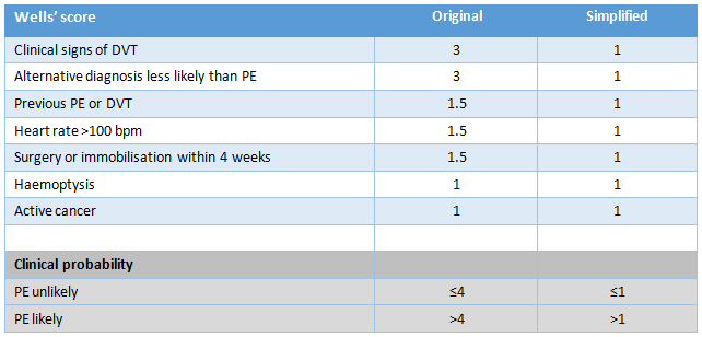 [Figure caption and citation for the preceding image starts]: Original and simplified Geneva score (revised)Created by the BMJ Knowledge Centre [Citation ends].
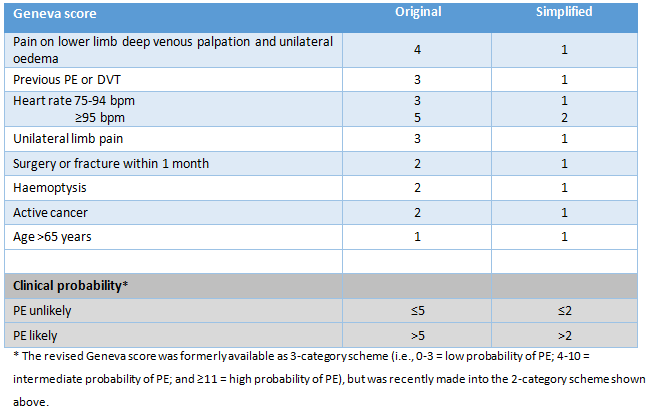 The simplified versions of the modified Wells Criteria or revised Geneva Score may be preferred in clinical practice because of their ease of use.[196]van Es N, Kraaijpoel N, Klok FA, et al. The original and simplified Wells rules and age-adjusted D-dimer testing to rule out pulmonary embolism: an individual patient data meta-analysis. J Thromb Haemost. 2017 Apr;15(4):678-84.
https://onlinelibrary.wiley.com/doi/full/10.1111/jth.13630
http://www.ncbi.nlm.nih.gov/pubmed/28106338?tool=bestpractice.com
Both simplified versions have been validated; neither has been shown to be superior to the other.[197]Hendriksen JM, Geersing GJ, Lucassen WA, et al. Diagnostic prediction models for suspected pulmonary embolism: systematic review and independent external validation in primary care. BMJ. 2015 Sep 8;351:h4438.
https://www.bmj.com/content/351/bmj.h4438.long
http://www.ncbi.nlm.nih.gov/pubmed/26349907?tool=bestpractice.com
However, the Geneva Score is based entirely on objective clinical items and may be more reproducible (the Wells Criteria [original and simplified] include the subjective clinical item 'alternative diagnosis less likely than PE').
The simplified versions of the modified Wells Criteria or revised Geneva Score may be preferred in clinical practice because of their ease of use.[196]van Es N, Kraaijpoel N, Klok FA, et al. The original and simplified Wells rules and age-adjusted D-dimer testing to rule out pulmonary embolism: an individual patient data meta-analysis. J Thromb Haemost. 2017 Apr;15(4):678-84.
https://onlinelibrary.wiley.com/doi/full/10.1111/jth.13630
http://www.ncbi.nlm.nih.gov/pubmed/28106338?tool=bestpractice.com
Both simplified versions have been validated; neither has been shown to be superior to the other.[197]Hendriksen JM, Geersing GJ, Lucassen WA, et al. Diagnostic prediction models for suspected pulmonary embolism: systematic review and independent external validation in primary care. BMJ. 2015 Sep 8;351:h4438.
https://www.bmj.com/content/351/bmj.h4438.long
http://www.ncbi.nlm.nih.gov/pubmed/26349907?tool=bestpractice.com
However, the Geneva Score is based entirely on objective clinical items and may be more reproducible (the Wells Criteria [original and simplified] include the subjective clinical item 'alternative diagnosis less likely than PE').
Investigations
Results of preliminary laboratory testing and radiographic investigations help to narrow the diagnosis. The choice of investigations is dictated by the clinical history and physical exam findings.
Pulse oximetry
Allows detection and ongoing monitoring of hypoxemia with initiation of oxygen supplementation as necessary, while undertaking diagnostic workup for its cause.
Methemoglobinaemia and carbon monoxide poisoning are not detectable with standard noninvasive pulse oximetry.
Peak expiratory flow (PEF)
Simple bedside test that may help differentiate between pulmonary and cardiac causes of dyspnea.
Low peak flow is associated with obstructive lung disease such as asthma, COPD, and cystic fibrosis.[198]Ailani RK, Ravakhah K, DiGiovine B, et al. Dyspnea differentiation index: a new method for the rapid separation of cardiac vs pulmonary dyspnea. Chest. 1999 Oct;116(4):1100-4.
http://www.ncbi.nlm.nih.gov/pubmed/10531178?tool=bestpractice.com
Arterial blood gas (ABG)
Not all patients with dyspnea display abnormal findings on ABG, and not all people with abnormal ABG results are dyspneic. However, the ABG results may help in constructing a differential diagnosis and, along with exertional oximetry, may be indicated to evaluate gas exchange abnormalities in conditions associated with hypoxemia.
Hypercapnia (PaCO₂ >45 mmHg) may accompany dyspnea in exacerbation of COPD, neuromuscular disease, upper airway obstruction, or obesity-hypoventilation syndrome.
Hypocapnia may be present in anxiety states and accompany any process that presents with hyperventilation, such as pulmonary embolism.
Hypoxemia (PaO₂ <70 mmHg at sea level) has a broad differential, including conditions causing shunting (acute respiratory distress syndrome, pneumonia, pulmonary edema, cyanotic valvular disease), ventilation-perfusion (V/Q) mismatching (COPD, asthma, pulmonary embolism), diffusion impairment (interstitial lung disease), or hypoventilation (COPD exacerbation, neuromuscular disease, upper airway obstruction, or obesity-hypoventilation syndrome).
The dyspnea differentiation index, which combines the PaO₂ with PEF ([PEF x PaO₂]/1000), has a reported diagnostic accuracy of 79% in differentiating cardiac from pulmonary causes of dyspnea.[198]Ailani RK, Ravakhah K, DiGiovine B, et al. Dyspnea differentiation index: a new method for the rapid separation of cardiac vs pulmonary dyspnea. Chest. 1999 Oct;116(4):1100-4.
http://www.ncbi.nlm.nih.gov/pubmed/10531178?tool=bestpractice.com
Acidosis (pH <7.36) is a potent stimulus of breathing and may accompany dyspnea in the late phases of almost any process presenting with dyspnea, including sepsis, pulmonary edema, exacerbation of COPD, renal failure, and cyanide toxicity.[199]Holland MA, Kozlowski LM. Clinical features and management of cyanide poisoning. Clin Pharm. 1986 Sep;5(9):737-41.
http://www.ncbi.nlm.nih.gov/pubmed/3530615?tool=bestpractice.com
It may also be present in idiopathic or medication-induced renal tubular acidosis and thiamine deficiency.[200]Ozawa H, Homma Y, Arisawa H, et al. Severe metabolic acidosis and heart failure due to thiamine deficiency. Nutrition. 2001 Apr;17(4):351-2.
http://www.ncbi.nlm.nih.gov/pubmed/11369178?tool=bestpractice.com
Acidosis may result from using medications such as nucleoside reverse-transcriptase inhibitors and topiramate.[153]Calza L, Manfredi R, Chiodo F. Hyperlactataemia and lactic acidosis in HIV-infected patients receiving antiretroviral therapy. Clin Nutr. 2005 Feb;24(1):5-15.
http://www.ncbi.nlm.nih.gov/pubmed/15681097?tool=bestpractice.com
[154]Delpirou-Nouh C, Gelisse P, Chanez P, et al. Migraine and topiramate induced dyspnea. Headache. 2007 Nov-Dec;47(10):1453-5.
http://www.ncbi.nlm.nih.gov/pubmed/17868349?tool=bestpractice.com
Alkalosis may be a consequence of anxiety, panic attacks, dehydration, pulmonary embolism, ovarian hyperstimulation syndrome, or pulmonary leukostasis.[201]Szyper-Kravitz M, Strahilevitz J, Oren V, et al. Pulmonary leukostasis: role of perfusion lung scan in diagnosis and follow up. Am J Hematol. 2001 Jun;67(2):136-8.
http://www.ncbi.nlm.nih.gov/pubmed/11343387?tool=bestpractice.com
[202]Abramov Y, Elchalal U, Schenker JG. Pulmonary manifestations of severe ovarian hyperstimulation syndrome: a multicenter study. Fertil Steril. 1999 Apr;71(4):645-51.
http://www.ncbi.nlm.nih.gov/pubmed/10202873?tool=bestpractice.com
Electrocardiogram (ECG)
Helps diagnose acute coronary syndromes as the cause of dyspnea with ST-T-segment changes. It also identifies complete heart block, bradycardias, and tachyarrhythmias, and detects changes suggestive of pericarditis, cardiac tamponade (low voltage), and pulmonary embolism.
Changes in the p-wave morphology may help diagnose right atrial enlargement (typical of a chronic pulmonary process) or left atrial enlargement (typical of valvular heart disease).
Change in the QRS axis may indicate right (COPD, pulmonary hypertension) or left (hypertension, valvular heart disease) ventricular enlargement or hypertrophy.
Spirometry
Simple office-based test allows the detection of an obstructive deficit, which is revealed by a disproportionate reduction in the forced expiratory volume in the first second of expiration (FEV1) in relation to the forced vital capacity (FVC).
Obstructive deficits are characteristic of asthma, emphysema, or chronic bronchitis.[24]Global Initiative for Asthma. Global strategy for asthma management and prevention. May 2024 [internet publication].
https://ginasthma.org/2024-report
[25]Global Initiative for Chronic Obstructive Lung Disease. Global strategy for prevention, diagnosis and management of COPD: 2025 report. 2024 [internet publication].
https://goldcopd.org/2025-gold-report
[203]Louis R, Satia I, Ojanguren I, et al. European Respiratory Society guidelines for the diagnosis of asthma in adults. Eur Respir J. 2022 Feb 15:2101585.
https://www.doi.org/10.1183/13993003.01585-2021
http://www.ncbi.nlm.nih.gov/pubmed/35169025?tool=bestpractice.com
More symmetric reduction in FEV1 and FVC may suggest restriction and warrants full pulmonary function testing, with measurement of lung volumes and DLCO (diffusing capacity of lung for carbon monoxide).[204]Dempsey TM, Scanlon PD. Pulmonary Function Tests for the Generalist: A Brief Review. Mayo Clin Proc. 2018 Jun;93(6):763-71.
http://www.ncbi.nlm.nih.gov/pubmed/29866281?tool=bestpractice.com
CBC
Leukocytosis may accompany dyspnea in any infectious process involving the respiratory system, as well as in sepsis, autoimmune disease, parasitic infections, and leukemia.[201]Szyper-Kravitz M, Strahilevitz J, Oren V, et al. Pulmonary leukostasis: role of perfusion lung scan in diagnosis and follow up. Am J Hematol. 2001 Jun;67(2):136-8.
http://www.ncbi.nlm.nih.gov/pubmed/11343387?tool=bestpractice.com
[205]Tsai HC, Lee SS, Liu YC, et al. Clinical manifestations of strongyloidiasis in southern Taiwan. J Microbiol Immunol Infect. 2002 Mar;35(1):29-36.
http://www.ncbi.nlm.nih.gov/pubmed/11950117?tool=bestpractice.com
Eosinophilia may be present in a dyspneic patient with parasitic disease, certain vasculitides (e.g., Churg-Strauss syndrome), asthma, eosinophilic pneumonia, drug reaction with eosinophilia and systemic symptoms (DRESS), or cocaine use.[205]Tsai HC, Lee SS, Liu YC, et al. Clinical manifestations of strongyloidiasis in southern Taiwan. J Microbiol Immunol Infect. 2002 Mar;35(1):29-36.
http://www.ncbi.nlm.nih.gov/pubmed/11950117?tool=bestpractice.com
[206]Martinez Alonso JC, Dominguez Ortega FJ, Fuentes Gonzalo MJ. Churg-Strauss syndrome in a case of eosinophilia. Allergol Immunopathol (Madr). 2004 Jul-Aug;32(4):238-40.
http://www.ncbi.nlm.nih.gov/pubmed/15324657?tool=bestpractice.com
[207]Tsapas A, Paletas K, Vlachaki E, et al. Eosinophilic pneumonia associated with heroin inhalation: a case report. Wien Klin Wochenschr. 2008;120(5-6):178-80.
http://www.ncbi.nlm.nih.gov/pubmed/18365158?tool=bestpractice.com
[208]Reyes F, Vaitkus V, Al-Ajam M. A case of cocaine-induced eosinophilic pneumonia: Case report and review of the literature. Respir Med Case Rep. 2018;23:98-102.
https://www.sciencedirect.com/science/article/pii/S2213007117303994
http://www.ncbi.nlm.nih.gov/pubmed/29487790?tool=bestpractice.com
[209]Taweesedt PT, Nordstrom CW, Stoeckel J, et al. Pulmonary manifestations of drug reaction with eosinophilia and systemic symptoms (DRESS) syndrome: a systematic review. Biomed Res Int. 2019 Sep 24;2019:7863815.
https://www.hindawi.com/journals/bmri/2019/7863815
http://www.ncbi.nlm.nih.gov/pubmed/31662996?tool=bestpractice.com
Anemia may be the primary reason for dyspnea or may accompany it in drug-related lung injury, hereditary hemorrhagic telangiectasia, acute chest syndrome of sickle cell disease, pulmonary alveolar hemorrhage, or widespread infectious processes.[210]Carloni A, Piciucchi S, Giannakakis K, et al. Diffuse alveolar hemorrhage after leflunomide therapy in a patient with rheumatoid arthritis. J Thorac Imaging. 2008 Feb;23(1):57-9.
http://www.ncbi.nlm.nih.gov/pubmed/18347524?tool=bestpractice.com
[211]Chauhan S, Singh R. Red spots and recurrent anemia. Am J Med. 2006 Sep;119(9):743-5.
http://www.ncbi.nlm.nih.gov/pubmed/16945607?tool=bestpractice.com
Thrombocytopenia may be present with dyspnea in viral infections, including influenza, SARS, and Hantavirus pulmonary syndrome.[86]Verity R, Prasad E, Grimsrud K, et al. Hantavirus pulmonary syndrome in northern Alberta, Canada: clinical and laboratory findings for 19 cases. Clin Infect Dis. 2000 Oct;31(4):942-6.
https://academic.oup.com/cid/article/31/4/942/376669/Hantavirus-Pulmonary-Syndrome-in-Northern-Alberta
http://www.ncbi.nlm.nih.gov/pubmed/11049774?tool=bestpractice.com
[87]Hui DS, Chan MC, Wu AK, et al. Severe acute respiratory syndrome (SARS): epidemiology and clinical features. Postgrad Med J. 2004 Jul;80(945):373-81.
http://pmj.bmj.com/content/80/945/373.long
http://www.ncbi.nlm.nih.gov/pubmed/15254300?tool=bestpractice.com
It may also be due to adverse drug reactions, especially with chemotherapy.
C-reactive protein
Electrolytes
Hyponatremia may accompany dyspnea in congestive heart failure, chronic kidney disease, liver failure, or hypothyroidism.
Thyroid function tests
Thyroid disease may present with dyspnea.[10]Ferry OR, Huang YC, Masel PJ, et al. Diagnostic approach to chronic dyspnoea in adults. J Thorac Dis. 2019 Oct;11(suppl 17):S2117-28.
https://www.ncbi.nlm.nih.gov/pmc/articles/PMC6831921
http://www.ncbi.nlm.nih.gov/pubmed/31737340?tool=bestpractice.com
Liver function tests
Bilirubin may be elevated in dyspneic patients with liver failure, congestive heart failure, leptospirosis, and thoracic amebiasis.[108]Vieira SR, Brauner JS. Leptospirosis as a cause of acute respiratory failure: clinical features and outcome in 35 critical care patients. Braz J Infect Dis. 2002 Jun;6(3):135-9.
http://www.scielo.br/scielo.php?script=sci_arttext&pid=S1413-86702002000300006&lng=en&nrm=iso&tlng=en
http://www.ncbi.nlm.nih.gov/pubmed/12144750?tool=bestpractice.com
[212]van Lingen R, Warshow U, Dalton HR, et al. Jaundice as a presentation of heart failure. J R Soc Med. 2005 Aug;98(8):357-9.
https://www.ncbi.nlm.nih.gov/pmc/articles/PMC1181835
http://www.ncbi.nlm.nih.gov/pubmed/16055900?tool=bestpractice.com
[213]Shamsuzzaman SM, Hashiguchi Y. Thoracic amebiasis. Clin Chest Med. 2002 Jun;23(2):479-92.
http://www.ncbi.nlm.nih.gov/pubmed/12092041?tool=bestpractice.com
Transaminases may be elevated in liver failure, acute myocardial infarction, atypical pneumonia (especially Legionella pneumonia), DRESS, and viral infections such as SARS and Hantavirus pulmonary syndrome.[86]Verity R, Prasad E, Grimsrud K, et al. Hantavirus pulmonary syndrome in northern Alberta, Canada: clinical and laboratory findings for 19 cases. Clin Infect Dis. 2000 Oct;31(4):942-6.
https://academic.oup.com/cid/article/31/4/942/376669/Hantavirus-Pulmonary-Syndrome-in-Northern-Alberta
http://www.ncbi.nlm.nih.gov/pubmed/11049774?tool=bestpractice.com
[87]Hui DS, Chan MC, Wu AK, et al. Severe acute respiratory syndrome (SARS): epidemiology and clinical features. Postgrad Med J. 2004 Jul;80(945):373-81.
http://pmj.bmj.com/content/80/945/373.long
http://www.ncbi.nlm.nih.gov/pubmed/15254300?tool=bestpractice.com
[209]Taweesedt PT, Nordstrom CW, Stoeckel J, et al. Pulmonary manifestations of drug reaction with eosinophilia and systemic symptoms (DRESS) syndrome: a systematic review. Biomed Res Int. 2019 Sep 24;2019:7863815.
https://www.hindawi.com/journals/bmri/2019/7863815
http://www.ncbi.nlm.nih.gov/pubmed/31662996?tool=bestpractice.com
Kidney function tests
Dyspnea accompanied by laboratory evidence of renal insufficiency may be due to metabolic acidosis, uremic pleurisy, long-term volume overload with pleural effusions, pneumonia, DRESS, and several types of acute vasculitis.[209]Taweesedt PT, Nordstrom CW, Stoeckel J, et al. Pulmonary manifestations of drug reaction with eosinophilia and systemic symptoms (DRESS) syndrome: a systematic review. Biomed Res Int. 2019 Sep 24;2019:7863815.
https://www.hindawi.com/journals/bmri/2019/7863815
http://www.ncbi.nlm.nih.gov/pubmed/31662996?tool=bestpractice.com
[214]Bakirci T, Sasak G, Ozturk S, et al. Pleural effusion in long-term hemodialysis patients. Transplant Proc. 2007 May;39(4):889-91.
http://www.ncbi.nlm.nih.gov/pubmed/17524841?tool=bestpractice.com
[215]Dreyer G, Fan S. Therapeutic implications of coexisting severe pulmonary hemorrhage and pulmonary emboli in a case of Wegener granulomatosis. Am J Kidney Dis. 2009 May;53(5):e5-8.
http://www.ncbi.nlm.nih.gov/pubmed/19303682?tool=bestpractice.com
[216]Green RJ, Ruoss SJ, Kraft SA, et al. Pulmonary capillaritis and alveolar hemorrhage: update on diagnosis and management. Chest. 1996 Nov;110(5):1305-16.
http://www.ncbi.nlm.nih.gov/pubmed/8915239?tool=bestpractice.com
Cardiac enzymes
Elevated troponin I/T, myoglobin, and creatine kinase-MB may accompany acute myocardial infarction, myocarditis, takotsubo cardiomyopathy, DRESS, or rarely hypothyroidism.[217]Buschmann IR, Bondke A, Elgeti T, et al. Positive cardiac troponin I and T and chest pain in a patient with iatrogenic hypothyroidism and no coronary artery disease. Int J Cardiol. 2007 Feb 7;115(2):e83-5.
http://www.ncbi.nlm.nih.gov/pubmed/17084920?tool=bestpractice.com
[218]Thygesen K, Alpert JS, Jaffe AS, et al. Fourth Universal Definition of Myocardial Infarction (2018). J Am Coll Cardiol. 2018 Oct 30;72(18):2231-64.
https://www.sciencedirect.com/science/article/pii/S0735109718369419
http://www.ncbi.nlm.nih.gov/pubmed/30153967?tool=bestpractice.com
[219]Husain Z, Reddy BY, Schwartz RA. DRESS syndrome: part I. clinical perspectives. J Am Acad Dermatol. 2013 May;68(5):693.e1-14.
http://www.ncbi.nlm.nih.gov/pubmed/23602182?tool=bestpractice.com
Troponin is the preferred biomarker in the evaluation of patients with suspected ACS. Myoglobin is a nonspecific marker, with an early peak in activity following myocardial injury. Other markers have satisfactory sensitivity if measured 4-6 hours following the onset of symptoms. Troponin I and T, and CK-MB, are specific to the heart muscle.
May also reflect chronic coronary artery disease with superimposed physiologic stress (e.g., sepsis, pulmonary embolism, chronic kidney disease).
BNP or N-terminal pro-BNP (NT-proBNP)
Elevated BNP and NT-proBNP have been associated with congestive heart failure, but also sepsis, coronary artery disease, pulmonary embolism, COPD with cor pulmonale, renal failure, liver cirrhosis, and hyperthyroidism.[220]Mant J, Doust J, Roalfe A, et al. Systematic review and individual patient data meta-analysis of diagnosis of heart failure, with modelling of implications of different diagnostic strategies in primary care. Health Technol Assess. 2009 Jul;13(32):1-207.
https://www.journalslibrary.nihr.ac.uk/hta/hta13320#/abstract
http://www.ncbi.nlm.nih.gov/pubmed/19586584?tool=bestpractice.com
[221]Worster A, Balion CM, Hill SA, et al. Diagnostic accuracy of BNP and NT-proBNP in patients presenting to acute care settings with dyspnea: a systematic review. Clin Biochem. 2008 Mar;41(4-5):250-9.
http://www.ncbi.nlm.nih.gov/pubmed/17915204?tool=bestpractice.com
[222]Lam LL, Cameron PA, Schneider HG, et al. Meta-analysis: effect of B-type natriuretic peptide testing on clinical outcomes in patients with acute dyspnea in the emergency setting. Ann Intern Med. 2010 Dec 7;153(11):728-35.
http://www.ncbi.nlm.nih.gov/pubmed/21135296?tool=bestpractice.com
[223]Trinquart L, Ray P, Riou B, et al. Natriuretic peptide testing in EDs for managing acute dyspnea: a meta-analysis. Am J Emerg Med. 2011 Sep;29(7):757-67.
http://www.ncbi.nlm.nih.gov/pubmed/20825895?tool=bestpractice.com
In emergency settings, natriuretic peptide biomarker levels may be more useful for ruling out than ruling in heart failure.[224]Heidenreich PA, Bozkurt B, Aguilar D, et al. 2022 AHA/ACC/HFSA guideline for the management of heart failure: a report of the American College of Cardiology/American Heart Association Joint Committee on clinical practice guidelines. Circulation. 2022 May 3;145(18):e895-1032.
https://www.ahajournals.org/doi/10.1161/CIR.0000000000001063
http://www.ncbi.nlm.nih.gov/pubmed/35363499?tool=bestpractice.com
A low normal BNP level (<100 nanograms/L [<100 picograms/mL]) or a low NT-proBNP level (<300 nanograms/L [<300 picograms/mL]) is helpful in excluding congestive heart failure.[66]National Institute for Health and Care Excellence. Acute heart failure: diagnosis and management. Nov 2021 [internet publication].
https://www.nice.org.uk/guidance/cg187
NT-proBNP levels <400 picograms/mL in an untreated person with chronic symptoms makes a diagnosis of heart failure less likely.[225]National Institute for Health and Care Excellence. Chronic heart failure in adults: diagnosis and management. September 2018 [internet publication].
https://www.nice.org.uk/guidance/ng106
D-dimer
D-dimer testing is particularly useful in patients with a low clinical probability assessment for PE.[49]Konstantinides SV, Meyer G, Becattini C, et al. 2019 ESC guidelines for the diagnosis and management of acute pulmonary embolism developed in collaboration with the European Respiratory Society (ERS): the Task Force for the diagnosis and management of acute pulmonary embolism of the European Society of Cardiology (ESC). Eur Respir J. 2019 Sep;543-603.
https://academic.oup.com/eurheartj/article/41/4/543/5556136
http://www.ncbi.nlm.nih.gov/pubmed/31504429?tool=bestpractice.com
[226]Crawford F, Andras A, Welch K, et al. D-dimer test for excluding the diagnosis of pulmonary embolism. Cochrane Database Syst Rev. 2016 Aug 5;2016(8):CD010864.
https://www.cochranelibrary.com/cdsr/doi/10.1002/14651858.CD010864.pub2/full
http://www.ncbi.nlm.nih.gov/pubmed/27494075?tool=bestpractice.com
If there is high clinical suspicion of PE, or the patient does not satisfy the PERC rule, the Wells (or Geneva) Score may be used to categorize the patient as:
“PE likely” (Wells Score >4); D-dimer testing is not recommended because a negative result will not remove the need for definitive imaging (usually computed tomographic pulmonary angiography [CTPA] if available)[55]National Institute for Health and Care Excellence. Venous thromboembolic diseases: diagnosis, management and thrombophilia testing. Aug 2023 [internet publication].
https://www.nice.org.uk/guidance/ng158
[227]Raja AS, Greenberg JO, Qaseem A, et al. Evaluation of patients with suspected acute pulmonary embolism: best practice advice from the clinical guidelines committee of the American College of Physicians. Ann Intern Med. 2015 Nov 3;163(9):701-11.
https://www.acpjournals.org/doi/10.7326/M14-1772
http://www.ncbi.nlm.nih.gov/pubmed/26414967?tool=bestpractice.com
[Figure caption and citation for the preceding image starts]: Original and simplified Wells Criteria (modified)Created by the BMJ Knowledge Centre [Citation ends].
“PE unlikely” (Wells Score ≤4); D-dimer test is ordered.[55]National Institute for Health and Care Excellence. Venous thromboembolic diseases: diagnosis, management and thrombophilia testing. Aug 2023 [internet publication].
https://www.nice.org.uk/guidance/ng158
[227]Raja AS, Greenberg JO, Qaseem A, et al. Evaluation of patients with suspected acute pulmonary embolism: best practice advice from the clinical guidelines committee of the American College of Physicians. Ann Intern Med. 2015 Nov 3;163(9):701-11.
https://www.acpjournals.org/doi/10.7326/M14-1772
http://www.ncbi.nlm.nih.gov/pubmed/26414967?tool=bestpractice.com
A normal plasma D-dimer level safely excludes PE in patients with an unlikely pretest probability of PE and no further investigation is required.[49]Konstantinides SV, Meyer G, Becattini C, et al. 2019 ESC guidelines for the diagnosis and management of acute pulmonary embolism developed in collaboration with the European Respiratory Society (ERS): the Task Force for the diagnosis and management of acute pulmonary embolism of the European Society of Cardiology (ESC). Eur Respir J. 2019 Sep;543-603.
https://academic.oup.com/eurheartj/article/41/4/543/5556136
http://www.ncbi.nlm.nih.gov/pubmed/31504429?tool=bestpractice.com
[227]Raja AS, Greenberg JO, Qaseem A, et al. Evaluation of patients with suspected acute pulmonary embolism: best practice advice from the clinical guidelines committee of the American College of Physicians. Ann Intern Med. 2015 Nov 3;163(9):701-11.
https://www.acpjournals.org/doi/10.7326/M14-1772
http://www.ncbi.nlm.nih.gov/pubmed/26414967?tool=bestpractice.com
D-dimer blood testing by enzyme-linked immunosorbent assay (ELISA) has a sensitivity and negative predictive value >95%, regardless of the calculated clinical probability, making it useful to rule out the presence of venous thromboembolism.[227]Raja AS, Greenberg JO, Qaseem A, et al. Evaluation of patients with suspected acute pulmonary embolism: best practice advice from the clinical guidelines committee of the American College of Physicians. Ann Intern Med. 2015 Nov 3;163(9):701-11.
https://www.acpjournals.org/doi/10.7326/M14-1772
http://www.ncbi.nlm.nih.gov/pubmed/26414967?tool=bestpractice.com
However, specificity is much lower, with estimates from 23% to 63%.[226]Crawford F, Andras A, Welch K, et al. D-dimer test for excluding the diagnosis of pulmonary embolism. Cochrane Database Syst Rev. 2016 Aug 5;2016(8):CD010864.
https://www.cochranelibrary.com/cdsr/doi/10.1002/14651858.CD010864.pub2/full
http://www.ncbi.nlm.nih.gov/pubmed/27494075?tool=bestpractice.com
Serologic tests
High titer antibody against the suspected antigen supports a diagnosis of hypersensitivity pneumonitis. Common causative antigens include bacteria (thermophilic Actinomycetes, responsible for farmer’s lung and mushroom picker’s lung) and animal proteins (responsible for pigeon breeder’s lung and bird fancier’s lung).
Blood cultures
Blood cultures should be obtained in patients with severe community acquired pneumonia (CAP). Severe CAP is defined by the presence of one major criterion.[37]Metlay JP, Waterer GW, Long AC, et al. Diagnosis and Treatment of Adults with Community-acquired Pneumonia. An Official Clinical Practice Guideline of the American Thoracic Society and Infectious Diseases Society of America. Am J Respir Crit Care Med. 2019 Oct 1;200(7):e45-e67.
https://www.atsjournals.org/doi/full/10.1164/rccm.201908-1581ST
http://www.ncbi.nlm.nih.gov/pubmed/31573350?tool=bestpractice.com
Septic shock with need for vasopressors
Respiratory failure requiring mechanical ventilation, or three or more minor criteria:[37]Metlay JP, Waterer GW, Long AC, et al. Diagnosis and Treatment of Adults with Community-acquired Pneumonia. An Official Clinical Practice Guideline of the American Thoracic Society and Infectious Diseases Society of America. Am J Respir Crit Care Med. 2019 Oct 1;200(7):e45-e67.
https://www.atsjournals.org/doi/full/10.1164/rccm.201908-1581ST
http://www.ncbi.nlm.nih.gov/pubmed/31573350?tool=bestpractice.com
Respiratory rate ≥30 breaths/min
PaO₂/FiO₂ ratio ≤ 250
Multilobar infiltrates
Confusion or disorientation
Uremia (blood urea nitrogen level ≥ 20 mg/dl)
Leukopenia (white blood cell count < 4,000 cells/μl)
Thrombocytopenia (platelet count < 100,000/μl)
Hypothermia (core temperature < 36°C)
Hypotension requiring aggressive fluid resuscitation
Blood cultures should also be obtained in patients who are being treated empirically for MRSA or P. aeruginosa; patients with previous P. aeruginosa or MRSA infection; and patients who have been hospitalized and received parenteral antibiotics within the last 90 days.[37]Metlay JP, Waterer GW, Long AC, et al. Diagnosis and Treatment of Adults with Community-acquired Pneumonia. An Official Clinical Practice Guideline of the American Thoracic Society and Infectious Diseases Society of America. Am J Respir Crit Care Med. 2019 Oct 1;200(7):e45-e67.
https://www.atsjournals.org/doi/full/10.1164/rccm.201908-1581ST
http://www.ncbi.nlm.nih.gov/pubmed/31573350?tool=bestpractice.com
Blood cultures have a yield of 2% to 9% in nonsevere community-acquired pneumonia (CAP) and are not recommended in these patients.[37]Metlay JP, Waterer GW, Long AC, et al. Diagnosis and Treatment of Adults with Community-acquired Pneumonia. An Official Clinical Practice Guideline of the American Thoracic Society and Infectious Diseases Society of America. Am J Respir Crit Care Med. 2019 Oct 1;200(7):e45-e67.
https://www.atsjournals.org/doi/full/10.1164/rccm.201908-1581ST
http://www.ncbi.nlm.nih.gov/pubmed/31573350?tool=bestpractice.com
Sputum cultures
Sputum Gram stain and culture is recommended for patients with severe community-acquired pneumonia; patients who are being treated empirically for MRSA or P. aeruginosa; patients with previous P. aeruginosa or MRSA infection; and patients who have been hospitalized and received parenteral antibiotics within the last 90 days.[37]Metlay JP, Waterer GW, Long AC, et al. Diagnosis and Treatment of Adults with Community-acquired Pneumonia. An Official Clinical Practice Guideline of the American Thoracic Society and Infectious Diseases Society of America. Am J Respir Crit Care Med. 2019 Oct 1;200(7):e45-e67.
https://www.atsjournals.org/doi/full/10.1164/rccm.201908-1581ST
http://www.ncbi.nlm.nih.gov/pubmed/31573350?tool=bestpractice.com
Urine pneumococcal antigen and Legionella antigen
Randomized trials have failed to identify a benefit for urinary antigen testing for S. pneumoniae and Legionella.[228]Falguera M, Ruiz-González A, Schoenenberger JA, et al. Prospective, randomised study to compare empirical treatment versus targeted treatment on the basis of the urine antigen results in hospitalised patients with community-acquired pneumonia. Thorax. 2010 Feb;65(2):101-6.
https://thorax.bmj.com/content/65/2/101.long
http://www.ncbi.nlm.nih.gov/pubmed/19703825?tool=bestpractice.com
There is concern that narrowing antibiotic therapy in response to positive urinary antigen tests could lead to increased risk of clinical relapse. Therefore urinary pneumococcal and Legionella antigen testing is only recommended for patients with severe CAP or if epidemiological factors increase risk of infection (e.g. recent travel, Legionella outbreak).[37]Metlay JP, Waterer GW, Long AC, et al. Diagnosis and Treatment of Adults with Community-acquired Pneumonia. An Official Clinical Practice Guideline of the American Thoracic Society and Infectious Diseases Society of America. Am J Respir Crit Care Med. 2019 Oct 1;200(7):e45-e67.
https://www.atsjournals.org/doi/full/10.1164/rccm.201908-1581ST
http://www.ncbi.nlm.nih.gov/pubmed/31573350?tool=bestpractice.com
Influenza virus tests
Influenza is a clinical diagnosis. The role of laboratory testing is to inform decisions about antiviral therapy, whether to perform other diagnostic testing, and the need to implement infection control measures. Not all patients with influenza require diagnostic testing.
Several available tests give results within 4 hours. These include molecular assays (rapid molecular assays; reverse transcription polymerase chain reaction [RT-PCR]) and antigen detection tests (rapid influenza diagnostic tests; immunofluorescence assays). Molecular assays are recommended for hospitalized patients with suspected influenza. They have a sensitivity of 90% to 95%.[229]Centers for Disease Control and Prevention (CDC). Influenza signs and symptoms and the role of laboratory diagnostics. Aug 2020 [internet publication].
https://www.cdc.gov/flu/professionals/diagnosis/overview-testing-methods.htm
A nasal or nasopharyngeal swab, wash, or aspirate should be collected as soon as possible after the onset of illness. Virus shedding in the upper respiratory tract declines significantly 3 to 4 days after illness onset.[230]Centers for Disease Control and Prevention. Manual for the surveillance of vaccine-preventable diseases: chapter 6 - influenza. Nov 2017 [internet publication].
https://www.cdc.gov/vaccines/pubs/surv-manual/chpt06-influenza.html
Viral culture takes 3 to 10 days so results do not inform clinical management, but are helpful for surveillance and antigenic characterization of new influenza strains.
Other respiratory viral testing
Nasopharyngeal secretions may be tested for respiratory syncytial virus (RSV), human metapneumovirus (hMPV), adenovirus, rhinovirus, and parainfluenza virus, by polymerase chain reaction (PCR).
Real-time RT-PCR for the detection of severe acute respiratory syndrome coronavirus 2 (SARS-CoV-2) viral RNA.
Chest x-ray
Appropriate for most adults with acute and chronic dyspnea.[9]American College of Radiology. ACR appropriateness criteria: dyspnea-suspected cardiac origin (ischaemia already excluded). 2021 [internet publication].
https:/acsearch.acr.org/docs/69407/Narrative
[11]American College of Radiology. ACR appropriateness criteria: chronic dyspnea - noncardiovascular origin. 2024 [internet publication].
https://acsearch.acr.org/docs/69448/Narrative
[231]Jokerst C, Chung JH, et al. Expert Panel on Thoracic Imaging. ACR Appropriateness Criteria® acute respiratory illness in immunocompetent patients. J Am Coll Radiol. 2018 Nov;15(suppl 11):S240-51.
https://www.jacr.org/article/S1546-1440(18)31154-2/fulltext
http://www.ncbi.nlm.nih.gov/pubmed/30392593?tool=bestpractice.com
Chest x-ray is important to exclude spontaneous or secondary pneumothorax.
Pulmonary venous congestion and an enlarged heart suggest congestive heart failure.[224]Heidenreich PA, Bozkurt B, Aguilar D, et al. 2022 AHA/ACC/HFSA guideline for the management of heart failure: a report of the American College of Cardiology/American Heart Association Joint Committee on clinical practice guidelines. Circulation. 2022 May 3;145(18):e895-1032.
https://www.ahajournals.org/doi/10.1161/CIR.0000000000001063
http://www.ncbi.nlm.nih.gov/pubmed/35363499?tool=bestpractice.com
An enlarged cardiac silhouette may indicate valvular heart disease, a pericardial cyst, or cardiac tamponade.[72]Roy CL, Minor MA, Brookhart MA, et al. Does this patient with a pericardial effusion have cardiac tamponade? JAMA. 2007 Apr 25;297(16):1810-8.
http://www.ncbi.nlm.nih.gov/pubmed/17456823?tool=bestpractice.com
[232]Patel J, Park C, Michaels J, et al. Pericardial cyst: case reports and a literature review. Echocardiography. 2004 Apr;21(3):269-72.
http://www.ncbi.nlm.nih.gov/pubmed/15053790?tool=bestpractice.com
Dyspnea accompanied by parenchymal infiltrates may be present in infectious pneumonia, pulmonary edema, eosinophilic pneumonitis, radiation pneumonitis, some interstitial lung diseases (sarcoidosis, usual interstitial pneumonitis, nonspecific interstitial pneumonitis, cryptogenic interstitial pneumonitis, lymphoid interstitial pneumonitis, acute interstitial pneumonitis, and pleuroparenchymal fibroelastosis), pneumoconioses (silicosis, asbestosis, berylliosis), drug-related lung disease, autoimmune disease-related lung disease (lupus, rheumatoid arthritis, scleroderma, polymyositis, vasculitis), metastatic lung disease and e-cigarette (or vaping product) use associated lung injury.[117]Evans ME, Twentyman E, Click ES, et al. Update: interim guidance for health care professionals evaluating and caring for patients with suspected e-cigarette, or vaping, product use-associated lung injury and for reducing the risk for rehospitalization and death following hospital discharge - United States, December 2019. MMWR Morb Mortal Wkly Rep. 2020 Jan 3;68(5152):1189-94.
https://www.ncbi.nlm.nih.gov/pmc/articles/PMC6943965
[233]Ghafoori P, Marks LB, Vujaskovic Z, et al. Radiation-induced lung injury: assessment, management, and prevention. Oncology (Williston Park). 2008 Jan;22(1):37-47.
http://www.ncbi.nlm.nih.gov/pubmed/18251282?tool=bestpractice.com
[234]Feinstein MB, DeSouza SA, Moreira AL, et al. A comparison of the pathological, clinical and radiographical, features of cryptogenic organising pneumonia, acute fibrinous and organising pneumonia and granulomatous organising pneumonia. J Clin Pathol. 2015 Jun;68(6):441-7.
https://jcp.bmj.com/content/68/6/441.long
http://www.ncbi.nlm.nih.gov/pubmed/25742910?tool=bestpractice.com
[235]Mathai SC, Danoff SK. Management of interstitial lung disease associated with connective tissue disease. BMJ. 2016 Feb 24;352:h6819.https://www.bmj.com/content/352/bmj.h6819.long
http://www.ncbi.nlm.nih.gov/pubmed/26912511?tool=bestpractice.com
[236]Baughman RP, Culver DA, Judson MA. A concise review of pulmonary sarcoidosis. Am J Respir Crit Care Med. 2011 Mar 1;183(5):573-81.
https://www.atsjournals.org/doi/full/10.1164/rccm.201006-0865CI
http://www.ncbi.nlm.nih.gov/pubmed/21037016?tool=bestpractice.com
[237]Orlandi M, Landini N, Bruni C, et al. Pleuroparenchymal fibroelastosis in rheumatic autoimmune diseases: a systematic literature review. Rheumatology (Oxford). 2020 Dec 1;59(12):3645-56.
https://academic.oup.com/rheumatology/article/59/12/3645/5904409
http://www.ncbi.nlm.nih.gov/pubmed/33313932?tool=bestpractice.com
Pleural effusion may accompany congestive heart failure, liver failure, uremia, nephrotic syndrome, malignancy, pneumonia, pulmonary embolism, or pleuritis.
Pleural thickening and nodularity may be seen in pleural tumors and pleuroparenchymal fibroelastosis.[85]Rena O, Filosso PL, Papalia E, et al. Solitary fibrous tumour of the pleura: surgical treatment. Eur J Cardiothorac Surg. 2001 Feb;19(2):185-9.
http://www.ncbi.nlm.nih.gov/pubmed/11167110?tool=bestpractice.com
[237]Orlandi M, Landini N, Bruni C, et al. Pleuroparenchymal fibroelastosis in rheumatic autoimmune diseases: a systematic literature review. Rheumatology (Oxford). 2020 Dec 1;59(12):3645-56.
https://academic.oup.com/rheumatology/article/59/12/3645/5904409
http://www.ncbi.nlm.nih.gov/pubmed/33313932?tool=bestpractice.com
Lung hyperinflation may be present in COPD, exacerbation of asthma, or foreign body aspiration.
Elevation of the hemidiaphragm may indicate its paralysis.
Unilateral lucidity may indicate pneumothorax or a diaphragmatic hernia.[238]Hanekamp LA, Toben FM. A pregnant woman with shortness of breath. Neth J Med. 2006 Mar;64(3):84, 95.
http://www.njmonline.nl/getpdf.php?id=400
http://www.ncbi.nlm.nih.gov/pubmed/16547361?tool=bestpractice.com
Prominent hilar vessels may be apparent in pulmonary hypertension.[239]American College of Radiology. ACR appropriateness criteria: suspected pulmonary hypertension, 2022 [internet publication].
https://www.acr.org/Clinical-Resources/ACR-Appropriateness-Criteria
Chest x-ray may not be needed in an acute exacerbation of asthma if there is no suspicion of pneumothorax or pneumonia.
Bedside ultrasonography of the chest
Point-of-care ultrasound (POCUS) exam of the heart, lungs, and inferior vena cava is an important tool in establishing the diagnosis of, or ruling out the presence of pleural effusion, pneumothorax, pulmonary edema, pneumonia, and pulmonary embolism.[240]Qaseem A, Etxeandia-Ikobaltzeta I, Mustafa RA, et al. Appropriate use of point-of-care ultrasonography in patients with acute dyspnea in emergency department or inpatient settings: a clinical guideline from the American College of Physicians. Ann Intern Med. 2021 Jul;174(7):985-93.
https://www.acpjournals.org/doi/10.7326/M20-7844
http://www.ncbi.nlm.nih.gov/pubmed/33900792?tool=bestpractice.com
[241]Gartlehner G, Wagner G, Affengruber L, et al. Point-of-care ultrasonography in patients with acute dyspnea: an evidence report for a clinical practice guideline by the American College of Physicians. Ann Intern Med. 2021 Jul;174(7):967-76.
https://www.acpjournals.org/doi/10.7326/M20-5504
http://www.ncbi.nlm.nih.gov/pubmed/33900798?tool=bestpractice.com
[242]Chan KK, Joo DA, McRae AD, et al. Chest ultrasonography versus supine chest radiography for diagnosis of pneumothorax in trauma patients in the emergency department. Cochrane Database Syst Rev. 2020 Jul 23;7(7):CD013031.
https://www.doi.org/10.1002/14651858.CD013031.pub2
http://www.ncbi.nlm.nih.gov/pubmed/32702777?tool=bestpractice.com
Presence of B-lines on bedside ultrasonography in a patient with high pretest probability of pulmonary edema is suggestive of pulmonary edema. Absence of B-lines in patients with low pretest probability of pulmonary edema almost rules out this diagnosis.[243]Al Deeb M, Barbic S, Featherstone R, et al. Point-of-care ultrasonography for the diagnosis of acute cardiogenic pulmonary edema in patients presenting with acute dyspnea: a systematic review and meta-analysis. Acad Emerg Med. 2014 Aug;21(8):843-52.
http://onlinelibrary.wiley.com/doi/10.1111/acem.12435/full
http://www.ncbi.nlm.nih.gov/pubmed/25176151?tool=bestpractice.com
Combining bedside echocardiography with ultrasonography of the chest may help further affirm the diagnosis of pulmonary edema.[244]Martindale JL, Wakai A, Collins SP, et al. Diagnosing acute heart failure in the emergency department: a systematic review and meta-analysis. Acad Emerg Med. 2016 Mar;23(3):223-42.
http://www.ncbi.nlm.nih.gov/pubmed/26910112?tool=bestpractice.com
Echocardiogram
Can detect pericardial disease and pulmonary hypertension.[239]American College of Radiology. ACR appropriateness criteria: suspected pulmonary hypertension, 2022 [internet publication].
https://www.acr.org/Clinical-Resources/ACR-Appropriateness-Criteria
[245]Humbert M, Kovacs G, Hoeper MM, et al. 2022 ESC/ERS guidelines for the diagnosis and treatment of pulmonary hypertension. Eur Heart J. 2022 Oct 11;43(38):3618-731.
https://academic.oup.com/eurheartj/article/43/38/3618/6673929
http://www.ncbi.nlm.nih.gov/pubmed/36017548?tool=bestpractice.com
May also be used to delineate valvular heart disease, measure diastolic dysfunction, and differentiate between systolic and diastolic failure.
The diagnosis of constrictive and restrictive heart diseases with heart failure requires echocardiographic study.[231]Jokerst C, Chung JH, et al. Expert Panel on Thoracic Imaging. ACR Appropriateness Criteria® acute respiratory illness in immunocompetent patients. J Am Coll Radiol. 2018 Nov;15(suppl 11):S240-51.
https://www.jacr.org/article/S1546-1440(18)31154-2/fulltext
http://www.ncbi.nlm.nih.gov/pubmed/30392593?tool=bestpractice.com
Computed tomographic (CT) angiography of the chest
CT pulmonary angiography confirms the diagnosis in most patients with suspicion of pulmonary embolism.
CT of the chest can also detect and help define the extent of pulmonary parenchymal disease (infectious and noninfectious infiltrates), suggest the presence of pulmonary edema, define airway abnormalities (benign stenoses, foreign body, malignancy), define vascular abnormalities (congenital and acquired stenoses of the intrathoracic blood vessels, aneurysms), confirm the presence of pleural effusion, or evaluate other intrathoracic structures (thymus, retrosternal goiter).
High resolution CT chest
Necessary in evaluating interstitial lung disease, which is not excluded by a normal chest x-ray.
Prone position imaging helps rule out processes that are gravity-dependent: for example, vascular congestion.
Expiratory imaging should be included in patients with obstructive and mixed pulmonary function tests to rule in or rule out air-trapping and tracheomalacia.[11]American College of Radiology. ACR appropriateness criteria: chronic dyspnea - noncardiovascular origin. 2024 [internet publication].
https://acsearch.acr.org/docs/69448/Narrative
[246]American College of Radiology. ACR appropriateness criteria: tracheobronchial Disease. 2024 [internet publication].
https://acsearch.acr.org/docs/3195161/Narrative
Ventilation-perfusion (V/Q) scan
Three-dimensional single-photon emission CT technology
Improves sensitivity and specificity of the V/Q scan.[247]Gutte H, Mortensen J, Jensen CV, et al. Detection of pulmonary embolism with combined ventilation-perfusion SPECT and low-dose CT: head-to-head comparison with multidetector CT angiography. J Nucl Med. 2009 Dec;50(12):1987-92.
http://jnm.snmjournals.org/content/50/12/1987.full
http://www.ncbi.nlm.nih.gov/pubmed/19910421?tool=bestpractice.com
Lateral x-ray of the neck
Chest fluoroscopy
CT coronary angiography
Coronary angiography is the conventional reference standard for diagnosis of coronary artery disease. Angiography also provides details about the overall coronary anatomy.
50% to 70% luminal diameter narrowing is considered coronary obstruction, although presence of lesser lesions is prognostically relevant.[248]Maddox TM, Stanislawski MA, Grunwald GK, et al. Nonobstructive coronary artery disease and risk of myocardial infarction. JAMA. 2014 Nov 5;312(17):1754-63.
https://www.ncbi.nlm.nih.gov/pmc/articles/PMC4893304
http://www.ncbi.nlm.nih.gov/pubmed/25369489?tool=bestpractice.com
Risks include thrombosis or hemorrhage related to vascular access, arrhythmia, and atheroembolism.
Pulmonary function testing
Involves spirometry, measuring lung volumes, and evaluating the DLCO.
The two most common patterns of ventilatory defect are an obstructive deficit (low FEV1/FVC ratio, increased residual volume, increased total lung capacity), seen in asthma, bronchitis, and emphysema, and a restrictive deficit (symmetric reduction of FEV1 and FVC, high FEV1/FVC ratio, low total lung capacity), seen in interstitial lung disease.
Less common patterns include a fixed or variable extrathoracic flow limitation in vocal cord obstruction and tumors, an isolated reduction of the DLCO in pulmonary hypertension and interstitial lung disease, and a low maximal voluntary ventilation in neuromuscular diseases.
Fractional exhaled nitric oxide testing
Measurement of a fraction of nitric oxide in exhaled air (FeNO, expressed in parts per billion [ppb]) can act as a noninvasive marker to detect eosinophilic inflammation of the airways. High FeNO level ( >40-50 ppb) supports the diagnosis of asthma.[203]Louis R, Satia I, Ojanguren I, et al. European Respiratory Society guidelines for the diagnosis of asthma in adults. Eur Respir J. 2022 Feb 15:2101585.
https://www.doi.org/10.1183/13993003.01585-2021
http://www.ncbi.nlm.nih.gov/pubmed/35169025?tool=bestpractice.com
[249]National Institute for Health and Care Excellence. Asthma: diagnosis, monitoring and chronic asthma management (BTS, NICE, SIGN). Nov 2024 [internet publication].
https://www.nice.org.uk/guidance/ng245
[250]Expert Panel Working Group of the National Heart, Lung, and Blood Institute (NHLBI) administered and coordinated National Asthma Education and Prevention Program Coordinating Committee (NAEPPCC)., Cloutier MM, Baptist AP, et al. 2020 Focused updates to the asthma management guidelines: a report from the National Asthma Education and Prevention Program Coordinating Committee Expert Panel Working Group. J Allergy Clin Immunol. 2020 Dec;146(6):1217-70.
https://www.doi.org/10.1016/j.jaci.2020.10.003
http://www.ncbi.nlm.nih.gov/pubmed/33280709?tool=bestpractice.com
Cutoff values may vary according to age and location; consult local guidance.
The National Heart, Lung, and Blood Institute (NHLBI) suggests FeNO measurement in individuals ages >5 years:[250]Expert Panel Working Group of the National Heart, Lung, and Blood Institute (NHLBI) administered and coordinated National Asthma Education and Prevention Program Coordinating Committee (NAEPPCC)., Cloutier MM, Baptist AP, et al. 2020 Focused updates to the asthma management guidelines: a report from the National Asthma Education and Prevention Program Coordinating Committee Expert Panel Working Group. J Allergy Clin Immunol. 2020 Dec;146(6):1217-70.
https://www.doi.org/10.1016/j.jaci.2020.10.003
http://www.ncbi.nlm.nih.gov/pubmed/33280709?tool=bestpractice.com
for whom the diagnosis of asthma is uncertain using history, examination, clinical course, spirometry, and bronchodilator responsiveness testing, or
in whom spirometry cannot be performed.
The UK National Institute for Health and Care Excellence guidelines on asthma recommend that adults, young people and children ages 5-16 years with a possible diagnosis of asthma should be offered a fractional exhaled nitric oxide (FeNO) test initially, before spirometry, if available.[249]National Institute for Health and Care Excellence. Asthma: diagnosis, monitoring and chronic asthma management (BTS, NICE, SIGN). Nov 2024 [internet publication].
https://www.nice.org.uk/guidance/ng245
The European Respiratory Society recommends that a FeNO test be offered to patients ages >18 years suspected of asthma, in whom the diagnosis has not been established based on the initial spirometry combined with bronchodilator reversibility testing.[203]Louis R, Satia I, Ojanguren I, et al. European Respiratory Society guidelines for the diagnosis of asthma in adults. Eur Respir J. 2022 Feb 15:2101585.
https://www.doi.org/10.1183/13993003.01585-2021
http://www.ncbi.nlm.nih.gov/pubmed/35169025?tool=bestpractice.com
Bronchial provocation test
Inhalation of increasing doses of methacholine or histamine can be used to establish the presence of bronchial hyperresponsiveness.
Bronchial hyperresponsiveness is a sensitive, but not specific, feature of asthma.[203]Louis R, Satia I, Ojanguren I, et al. European Respiratory Society guidelines for the diagnosis of asthma in adults. Eur Respir J. 2022 Feb 15:2101585.
https://www.doi.org/10.1183/13993003.01585-2021
http://www.ncbi.nlm.nih.gov/pubmed/35169025?tool=bestpractice.com
Nocturnal oximetry
Holter monitoring
Cardiopulmonary exercise testing
Involves a detailed analysis of the cardiorespiratory response to exercise and allows the evaluation of cardiac function, pulmonary gas exchange, and ventilation, and the detection of cardiac ischemia, exercise-related obstructive lung disease, and deconditioning.[251]Guazzi M, Bandera F, Ozemek C, et al. Cardiopulmonary exercise testing: what is its value? J Am Coll Cardiol. 2017 Sep 26;70(13):1618-36.
https://www.sciencedirect.com/science/article/pii/S0735109717392501
http://www.ncbi.nlm.nih.gov/pubmed/28935040?tool=bestpractice.com
Subjective intensity of dyspnea correlates closely with the magnitude and duration of respiratory effort during exertion.[252]Laveneziana P, Agostoni P. Exertional dyspnoea in cardiorespiratory disorders: the clinical use of cardiopulmonary exercise testing. Eur Respir Rev. 2016 Sep;25(141):227-9.
https://err.ersjournals.com/content/25/141/227.long
http://www.ncbi.nlm.nih.gov/pubmed/27581822?tool=bestpractice.com
Electromyography
Signs of acute or chronic denervation or reinnervation may accompany amyotrophic lateral sclerosis.[253]Hardiman O, Al-Chalabi A, Chio A, et al. Amyotrophic lateral sclerosis. Nat Rev Dis Primers. 2017 Oct 5;3:17071.
https://www.nature.com/articles/nrdp201771
http://www.ncbi.nlm.nih.gov/pubmed/28980624?tool=bestpractice.com
Lung biopsy
Thoracoscopic lung biopsy may be used to diagnose interstitial lung diseases. Lung biopsy may not be necessary if there are typical clinical and radiologic findings.
May also be used in diagnosing autoimmune or vascular diseases of the lung.
Transthoracic needle aspiration may be useful in confirming the malignant etiology of intrathoracic nodules.
Coronavirus disease 2019 (COVID-19)
Cough, fever, and dyspnea are the most common symptoms of COVID-19. The cough is usually dry. Other symptoms include anosmia, ageusia, fatigue, anorexia, myalgia, and sore throat. There may be a travel history to an affected area or close contact with a suspected or confirmed case in the 14 days prior to symptom onset. The median time from onset of symptoms to hospital admission is reported to be approximately 7 days.[258]Huang C, Wang Y, Li X, et al. Clinical features of patients infected with 2019 novel coronavirus in Wuhan, China. Lancet. 2020 Feb 15;395(10223):497-506.
https://www.thelancet.com/journals/lancet/article/PIIS0140-6736(20)30183-5/fulltext
http://www.ncbi.nlm.nih.gov/pubmed/31986264?tool=bestpractice.com
Physical exam may detect fever (with or without chills/rigors) and obvious cough and/or difficulty breathing. Auscultation of the chest may reveal inspiratory crackles, rales, and/or bronchial breathing in patients with pneumonia or respiratory distress. Patients with respiratory distress may have tachycardia, tachypnea, or cyanosis accompanying hypoxia.
The most common laboratory abnormalities in patients hospitalized with pneumonia include leukopenia, lymphopenia, leukocytosis, elevated liver transaminases, elevated lactate dehydrogenase, and elevated C-reactive protein. Other abnormalities include neutrophilia, thrombocytopenia, decreased hemoglobin, decreased albumin, and renal impairment.[258]Huang C, Wang Y, Li X, et al. Clinical features of patients infected with 2019 novel coronavirus in Wuhan, China. Lancet. 2020 Feb 15;395(10223):497-506.
https://www.thelancet.com/journals/lancet/article/PIIS0140-6736(20)30183-5/fulltext
http://www.ncbi.nlm.nih.gov/pubmed/31986264?tool=bestpractice.com
[259]Chen N, Zhou M, Dong X, et al. Epidemiological and clinical characteristics of 99 cases of 2019 novel coronavirus pneumonia in Wuhan, China: a descriptive study. Lancet. 2020 Feb 15;395(10223):507-13.
https://www.thelancet.com/journals/lancet/article/PIIS0140-6736(20)30211-7/fulltext
http://www.ncbi.nlm.nih.gov/pubmed/32007143?tool=bestpractice.com
[260]Wang D, Hu B, Hu C, et al. Clinical characteristics of 138 hospitalized patients with 2019 novel coronavirus-infected pneumonia in Wuhan, China. JAMA. 2020 Feb 7 [Epub ahead of print].
https://jamanetwork.com/journals/jama/fullarticle/2761044
http://www.ncbi.nlm.nih.gov/pubmed/32031570?tool=bestpractice.com
[261]Li LQ, Huang T, Wang YQ, et al. 2019 novel coronavirus patients' clinical characteristics, discharge rate and fatality rate of meta-analysis. J Med Virol. 2020 Mar 12 [Epub ahead of print].
https://onlinelibrary.wiley.com/doi/epdf/10.1002/jmv.25757
http://www.ncbi.nlm.nih.gov/pubmed/32162702?tool=bestpractice.com
[262]Qin C, Zhou L, Hu Z, et al. Dysregulation of immune response in patients with COVID-19 in Wuhan, China. Clin Infect Dis. 2020 Mar 12 [Epub ahead of print].
https://academic.oup.com/cid/advance-article/doi/10.1093/cid/ciaa248/5803306
http://www.ncbi.nlm.nih.gov/pubmed/32161940?tool=bestpractice.com
Blood and sputum specimens should be collected for culture in all patients to rule out other causes of lower respiratory tract infection and sepsis, especially in patients with an atypical epidemiologic history. Specimens should be collected prior to starting empiric antimicrobials if possible.
Molecular testing is required to confirm the diagnosis. Diagnostic tests should be performed according to guidance issued by local health authorities and should adhere to appropriate biosafety practices.
A chest x-ray should be ordered in all patients with suspected pneumonia. Unilateral lung infiltrates are found in 25% of patients, and bilateral lung infiltrates are found in 75% of patients. [258]Huang C, Wang Y, Li X, et al. Clinical features of patients infected with 2019 novel coronavirus in Wuhan, China. Lancet. 2020 Feb 15;395(10223):497-506.
https://www.thelancet.com/journals/lancet/article/PIIS0140-6736(20)30183-5/fulltext
http://www.ncbi.nlm.nih.gov/pubmed/31986264?tool=bestpractice.com
[259]Chen N, Zhou M, Dong X, et al. Epidemiological and clinical characteristics of 99 cases of 2019 novel coronavirus pneumonia in Wuhan, China: a descriptive study. Lancet. 2020 Feb 15;395(10223):507-13.
https://www.thelancet.com/journals/lancet/article/PIIS0140-6736(20)30211-7/fulltext
http://www.ncbi.nlm.nih.gov/pubmed/32007143?tool=bestpractice.com
[263]Song F, Shi N, Shan F, et al. Emerging coronavirus 2019-nCoV pneumonia. Radiology. 2020 Feb 6;200274.
https://pubs.rsna.org/doi/10.1148/radiol.2020200274
http://www.ncbi.nlm.nih.gov/pubmed/32027573?tool=bestpractice.com
All imaging procedures should be performed according to local infection prevention and control procedures to prevent transmission.
See our Coronavirus disease 2019 (COVID-19) topic for current diagnostic advice.