Differentials
Melanoma
SIGNS / SYMPTOMS
Tend to be >6 mm, more irregularly bordered, more asymmetric, haphazardly multicolored, with pink, red, white, brown, black, and blue components.[5] Referral to a dermatologist is encouraged if the clinician has any doubts in distinguishing a nevus from a melanoma. Excluding malignant change in nevi or de novo melanoma may be challenging, especially when distinguishing from dysplastic or Clark nevi.
INVESTIGATIONS
Dermatoscopy: ramified streaks of asymmetric pigment, black pepper-grain dots, bulbar pseudopods, blue-whitish veils, depigmented areas, multicolored pattern, milky red globules, irregular hairpin vessels, ulceration, asymmetric follicular openings, and rhomboid structures on the face.[37][Figure caption and citation for the preceding image starts]: Dermatoscopy of melanomaFrom the collection of Laurel Schwartz, Thomas Jefferson University [Citation ends].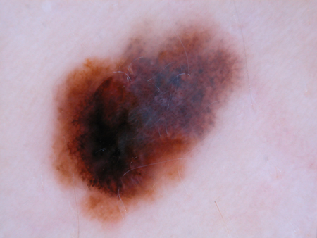
Histopathology: loss of organized nested melanocytes, and an increase in single melanocytes, often in a pagetoid pattern in the epidermis, with pleomorphism, cytologic atypia, and mitotic figures.
Seborrheic keratosis
SIGNS / SYMPTOMS
Waxy, "stuck-on" papules, often multiple, that increase in number over the age of 30 years.
INVESTIGATIONS
Dermatoscopy: comedo-like follicular pseudo-openings; horny pseudocysts, fissures, and crypts, fingerprint-like patterns, uniform hairpin vessels, and pigment pseudo network on the face.[37]
Histopathology can also distinguish.
Solar lentigo
SIGNS / SYMPTOMS
Flat, brown macules and patches on sun-damaged skin.
INVESTIGATIONS
Dermatoscopy: moth-eaten borders with an even brown reticular pattern.
Histopathology can also distinguish.
Simple lentigo
SIGNS / SYMPTOMS
Small, flat, brown macules clinically very similar to junctional nevi, found on nonsun-damaged skin.
INVESTIGATIONS
Dermatoscopy: small, evenly pigmented, brown, reticular pattern.
Histopathology can also distinguish.
Ephelis (freckle)
SIGNS / SYMPTOMS
Multiple flat, light-brown, sun-exposed macules, usually on the face, upper back, dorsal arms, and chest.
INVESTIGATIONS
History and physical exam can usually distinguish.
Dermatoscopy: similar to a solar lentigo but smaller in size.
Histopathology can also distinguish.
Cafe-au-lait macule
SIGNS / SYMPTOMS
Small-to-large, flat, brown homogeneous macules or patches with a well-defined and regular border, present since birth and unchanged, with exception of growth proportional to the person.
INVESTIGATIONS
History and physical exam can usually distinguish.
Histopathology can also distinguish.
Pigmented basal cell carcinoma (BCC)
SIGNS / SYMPTOMS
Arising on sun-exposed skin, the pigmented variant of BCC has irregular blue-gray pigment, tends to be asymmetric, and may be ill-defined.
INVESTIGATIONS
Dermatoscopy: structureless areas; maple leaf-type structures; branchlike telangiectasias; large, ovoid, blue-grayish nests; radial areas; and ulceration.[37]
Histopathology can also distinguish.
Dermatofibroma
SIGNS / SYMPTOMS
A firm, dome-shaped, often hyperpigmented or pink papule, often on the lower extremities of young adults, and thought to be induced by trauma (e.g., bug-bite).
INVESTIGATIONS
Dermatoscopy: central white, scarlike area with pigment at the periphery.
Histopathology can also distinguish.
Hemangioma
SIGNS / SYMPTOMS
A red-to-purple, dome-shaped, soft papule.
INVESTIGATIONS
Dermatoscopy: blood-filled, red-blue lacunae.
Histopathology can also distinguish.
Becker nevus
SIGNS / SYMPTOMS
Large brown patch usually on the upper back or chest, which often becomes darker and hairy after puberty. More common in men.
INVESTIGATIONS
Physical exam can usually distinguish.
Use of this content is subject to our disclaimer