Tests
1st tests to order
pulse oximetry
Test
At sea level, capillary blood oxygen saturations (SpO₂) of ≥90% are considered normal. Acute drops in SpO₂ <80% are associated with respiratory failure. Generally, when SpO₂ is <92% at sea level in a patient showing clinical signs of acute respiratory failure, an arterial blood gas will be helpful in evaluating and managing the case.
When available, continuous monitoring is important.
Along with SpO₂ saturation measurements, serum bicarbonate (HCO₃) may help in determining if underlying COPD with carbon dioxide retention is present (elevated HCO₂) or if acidosis is present (low HCO₃).
Inaccuracies can occur with poor fingertip perfusion (where monitor probes are usually attached). This problem may be overcome by attaching the probe to the ear lobe.
Nail polish, darkly pigmented skin, anemia, motion artifact, bright lighting in the area, carboxyhemoglobinemia, and methemoglobinemia can also reduce accuracy.[27][40]
Result
SpO₂ <80%
arterial blood gases
Test
Normal arterial partial pressure of oxygen (PaO₂) is >90 mmHg (>12 kPa) at sea level, on room air.
Normal arterial partial pressure of carbon dioxide (PaCO₂) is 35 to 45mmHg (4.7 to 6.0 kPa) at sea level, on room air. Normal pH is 7.38 to 7.42.
Analysis of arterial blood gas sampling allows classification into hypoxemic, hypercapnic, and mixed categories.
Result
pH <7.38; PaO₂ <60 mmHg (<8 kPa) (or <50 mmHg [<6.7 kPa] in chronic lung disease) on room air; PaCO₂ >50 mmHg (>6.7 kPa) on room air
Tests to consider
CBC
Test
An elevated or low white blood cell count can indicate the presence of infection. A change in the white blood cell profile to more leukocytes and immature cell forms can indicate infection.
Result
elevated white blood cell count
D-dimer
Test
Normal range or nonelevated D-dimer may help rule out acute pulmonary embolism.
D-dimer testing is limited because of the potential for significant false positives.[30]
Result
elevated D-dimer may indicate pulmonary thromboembolism; nonelevated D-dimer may exclude pulmonary thromboembolism
serum bicarbonate (HCO₃)
Test
Commonly reported as part of the serum electrolytes profile. Chronic respiratory acidosis, commonly seen with COPD, is associated with raised HCO₃, which slowly develops over time. A raised HCO₃ level, in association with developing acute respiratory failure implies underlying COPD as a comorbid condition.[29]
Result
may be elevated
Cardiac troponin I and/or T
Test
In addition to obtaining an ECG, cardiac specific troponin is recommended.
Raised cardiac specific troponin requires further cardiac evaluation, often with cardiothoracic ultrasound to directly assess myocardial activity and strain.
Result
raised troponin will alert the clinician to potential acute myocardial damage or disease or cardiac strain as found with acute pulmonary embolism
ECG
Test
Pulmonary embolism, which can lead to acute respiratory failure, may cause sinus tachycardia, atrial fibrillation, and/or evidence of myocardial ischemia. It is uncommon, but there may also be right bundle branch block, right axis deviation, and an S wave in lead I, a Q wave in lead III, and an inverted T wave in lead III (known as the S1Q3T3 pattern).
The ECG may also be useful in determining if cardiac ischemia or myocardial infarction is present. Acute coronary syndrome caused by cardiac ischemia or infarct can lead to acute respiratory failure and be further complicated with cardiogenic shock.
There may also be evidence of heart disease in patients with cardiac causes of acute respiratory failure, particularly in the setting of acute pulmonary edema or decompensated congestive heart failure. Emergency management of tachycardia or bradycardia will often improve cardiac output and consequently respiratory status.
Result
variable
chest x-ray
Test
Diffuse or patchy infiltrates on chest x-ray are associated with pneumonia, pulmonary edema, aspiration, progressive interstitial lung disease, pulmonary contusion, and alveolar hemorrhage.
Minimal changes in the chest x-ray are often seen in acute exacerbations of COPD, asthma, pulmonary embolism, and respiratory depression.
A chest x-ray is also used to assess for pneumothorax, collapse, and pulmonary effusions.
In asthma, hyperinflation of the lungs is associated with severe small airways obstruction.
In children, asymmetry of lucency of the lungs suggests foreign body aspiration.
Result
diffuse or patchy infiltrates; pneumothorax; pulmonary effusion; hyperinflation; asymmetric opacification of lung fields; asymmetric lucency of lung fields
pulmonary function tests
Test
If the peak expiratory flow rate (PEFR) or forced expiratory volume (FEV) is <35% to 50% of predicted for age, height, weight, and sex, then these can be predictive of respiratory compromise and potential acute respiratory failure. Monitoring the trend in these pulmonary function tests provides clinical evidence of improvement or deterioration in respiratory function.

How to use a peak flow meter to obtain a peak expiratory flow measurement.
An FEV in 1 second (FEV1) of <50% of sex- and age-predicted value indicates severe respiratory impairment.[44]
Negative inspiratory force (NIF) values above -25 cm H₂O (i.e., less negative flow) are associated with respiratory failure.[5]
Result
PEFR <35% to 50% of predicted; FEV <35% to 50% of predicted; FVC <50% to 70% of predicted; FEV1 <50% of predicted; NIF above -25 cm H₂O
Urine or serum toxicology
Test
Toxicology testing of serum and urine can be helpful in confirming suspected medication or illicit drug causes of respiratory failure (e.g., opioid overdose).
Result
identification of select drugs of abuse (available results will vary depending on the drug screen used)
chest CT
Test
CT can reveal chronic lung disease, pulmonary consolidation and effusion, parenchymal disease, bronchiectasis, and pulmonary embolism involving large- and medium-sized pulmonary arteries.
Result
pulmonary embolism (involving large- and medium-sized pulmonary arteries); chronic lung disease; pulmonary consolidation and effusion; parenchymal disease; bronchiectasis
CT pulmonary angiography (CTPA)
Test
CTPA is the imaging study of choice in patients with an abnormal D-dimer or a high probability of pulmonary embolism.[32] A CTPA uses contrast medium, which is administered intravenously at the same time as the CT scan of the chest. This allows filling defects to be visualized in the segmental and subsegmental branches of the pulmonary circulation. It has the highest diagnostic accuracy for pulmonary embolism of all the advanced noninvasive imaging methods.[32]
A CTPA is contraindicated in patients who have an allergy to contrast media or have renal failure.[33] It should also be avoided in pregnant women.[34]
Result
pulmonary embolism (involving large- and medium-sized pulmonary arteries); chronic lung disease; pulmonary consolidation and effusion; parenchymal disease; bronchiectasis
ventilation/perfusion lung scan
Test
V/Q lung scan, preferably using single photon emission computed tomography (SPECT, which may reduce the number of inconclusive scans), is an alternative to CT pulmonary angiography (CTPA).[35]
Result
pulmonary embolism (PE) likely when an area of ventilation is not perfused; a negative V/Q scan effectively excludes PE
capnometry
Test
Side-stream capnography can be used when patients are not intubated and is becoming more commonly used in assessment and monitoring of patients with acute illness or injury, to detect worsening respiratory condition.
Result
abnormally low or high expired CO₂
cardiothoracic ultrasound
Test
A complementary diagnostic tool that contributes to an early therapeutic decision based on reproducible pathophysiologic data. It is a noninvasive bedside test that can be used to initiate therapy pending confirmation by more advanced imaging techniques. Thoracic ultrasound is also of use in directing needle location for thoracentesis (drainage) procedures and placement of drainage devices.[36][37]
Result
evidence of effusion, pneumothorax, consolidation, or abscess; abnormal right ventricular filling or distension
Emerging tests
transcutaneous CO₂ monitoring
Test
Has a potential use in continuously monitoring the PaCO₂ of patients who are being maintained with noninvasive ventilatory support.[38] At present, transcutaneous CO₂ monitoring technology is developing and current agreement between PaCO₂ and transcutaneous PCO₂ has been questioned as being suboptimal.[39]

How to obtain an arterial blood sample from the radial artery.
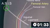
How to perform a femoral artery puncture to collect a sample of arterial blood.
Result
reduced arterial partial pressure of carbon dioxide (PaCO₂)
Use of this content is subject to our disclaimer