Images and videos
Images
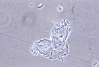


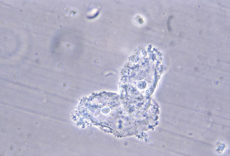
Vaginitis
Phase contrast wet mount micrograph of a vaginal discharge revealing the presence of Trichomonas vaginalis protozoa
CDC Image Library
See this image in context in the following section/s:

Vaginitis
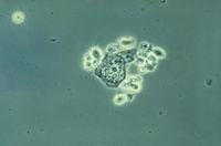
Vaginal smear identifying Candida albicans using a wet mount technique
CDC Image Library; Dr Stuart Brown
See this image in context in the following section/s:

Vaginitis
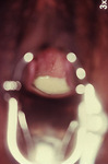
Trichomonas vaginitis with copious purulent discharge emanating from the cervical os
CDC Image Library
See this image in context in the following section/s:
Vaginitis
Photomicrograph revealing bacteria adhering to vaginal epithelial cells, known as clue cells
CDC Image Library; M. Rein
See this image in context in the following section/s:
Use of this content is subject to our disclaimer