History and exam
Key diagnostic factors
common
positive family history of epidermolysis bullosa (EB)
The only known risk factor for EB is a positive family history. Most patients with confirmed autosomal dominant forms of EB have a family history of the disease occurring in a parent.
mechanical fragility of the skin
With the rarest of exceptions, patients with EB will have a history of mechanically fragile skin.
If minor traction is applied to normal-appearing skin, a fresh peel (Nikolsky sign) can be induced.
recurrent blisters and erosions
A characteristic feature of all forms of EB. [Figure caption and citation for the preceding image starts]: Extensive blistering on leg, foot, and hand of an infant with EBS, generalized severe (EBS-gen sev)From the collection of J.D. Fine, MD [Citation ends].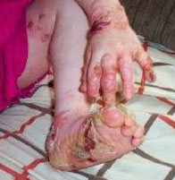
poorly healing wounds
Complex, with multiple potential contributory factors in addition to underlying skin fragility, including: persistent inflammation; microbial colonization and infection; anemia; fibrosis; scarring; and altered protease activity.
onset of cutaneous signs at birth or early infancy
When widespread blistering occurs at this early age, one of the more generalized subtypes of EB is more likely.
Some of the extracutaneous complications may not occur until late childhood or adulthood.
resolution of blistering within the first 1-2 years of life
Characteristic of bullous dermolysis of the newborn, a rare subtype of both types of dystrophic EB.[34]
generalized or localized distribution of skin involvement
Most common pattern of distribution for EB subtypes is either generalized or localized (e.g., palms and soles in localized EB simplex [EBS-loc]).
combination of milia, scarring, and dystrophic nails
Although all of the major EB types and subtypes overlap considerably, this combination of signs is most suggestive of dystrophic EB (DEB).
absence of milia, scarring, and dystrophic nails
Most consistent with EB simplex (EBS).[39]
exuberant granulation tissue
Nearly pathognomonic of severe junctional EB (JEB).[39][Figure caption and citation for the preceding image starts]: Axillary contractions and granulation tissue in a child with junctional EB, generalized severe (JEB-gen sev)From the collection of J.D. Fine, MD [Citation ends].
herpetiform blistering
Polycyclic or arcuate array of blisters or vesicles is pathognomonic of severe EBS.[39]
pseudosyndactyly
Most commonly occurs in recessive dystrophic EB (RDEB), especially severe RDEB. [Figure caption and citation for the preceding image starts]: Hand deformities, extensive scarring of wrists and legs in a child with recessive dystrophic EB, generalized severe (RDEB-gen sev)From the collection of J.D. Fine, MD [Citation ends].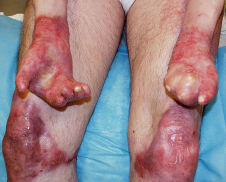
May also arise in all other RDEB subtypes, dominant dystrophic EB (DDEB), and rarely even intermediate junctional EB.[40][41]
enamel hypoplasia
Seen only in junctional EB (JEB) and occurs in all patients with JEB.[42]
reticulate hyperpigmentation
Characteristic of EB simplex with mottled pigmentation (EBS-MP).
muscular dystrophy
Associated with the subtype EB simplex with muscular dystrophy (EBS-MD). Of importance, this complication may not occur until young adulthood in some patients.
tracheolaryngeal stenosis or stricture
Occurs almost exclusively in junctional EB (JEB), most commonly severe JEB.[43]
severe upper airway disease
Associated with both generalized junctional EB (JEB) and JEB, laryngo-onycho-cutaneous syndrome (JEB-LOC).
uncommon
onset in mid or late childhood
More indicative of localized EB simplex (EBS-loc), or late-onset junctional EB (JEB-lo).
inverse (intertriginous), acral, or centripetal distribution of skin involvement
These distributions are rare, but when they are present they are pathognomonic for specific EB subtypes: recessive dystrophic EB, inversa (RDEB-I); junctional EB, inversa (JEB-I); and RDEB, centripetalis (RDEB-Ce).
These patterns usually only become more apparent after the first year of life.
Distribution types have specific prognostic implications.
Risk factors
strong
Family history of EB
Family history of EB is the only known risk factor for this disease.
Although spontaneous mutations do arise among autosomal dominant (AD) forms of EB, most patients have a positive family history of the disease occurring in a parent.
Of those who do not have a positive family history, particularly those with dystrophic EB, subsequent mutational analyses have demonstrated autosomal recessive (AR) rather than AD transmission in most.
Incomplete penetrance among AD forms of EB is extremely rare.
The risk to an affected parent heterozygous for AD EB of having an affected child is 50% with each pregnancy; all other children will be unaffected.
By definition, each parent of a child with AR EB is a silent carrier for the causative mutation. With each additional pregnancy, the risk of having an affected child, a silent carrier, or a genetically unaffected child is 25%, 50%, or 25%, respectively.
If a patient with an AR form of EB, who is, by definition, homozygous or compound heterozygous for the mutation, chooses to have children and has a genetically unaffected mate, then all of their offspring will be silent carriers for the disease.
Use of this content is subject to our disclaimer