Images and videos
Images


Abusive head trauma in infants and young children
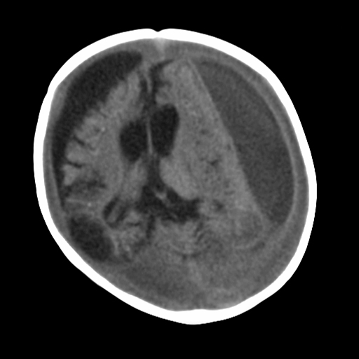
CT scan revealing subdural hemorrhage extending over the right convexity and in the intrahemispheric region, as well as enlargement of the extra-axial fluid spaces
From the personal collection of Alice Newton, MD; used with permission
See this image in context in the following section/s:

Abusive head trauma in infants and young children
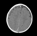
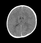
CT findings in fatal abusive head trauma often reveal significant brain edema with loss of gray-white differentiation and effacement of the ventricles. Subdural blood is often difficult to appreciate in such cases
From the personal collection of Alice Newton, MD; used with permission
See this image in context in the following section/s:
Abusive head trauma in infants and young children
MRI depicting subdural hygromas surrounding severe brain atrophy from abusive head trauma. This child was initially erroneously diagnosed with meningitis
From the personal collection of Alice Newton, MD; used with permission
See this image in context in the following section/s:

Abusive head trauma in infants and young children
Bruising on the ear of a 10-month-old infant
Reproduced with permission from Backhouse L et al. Unexplained bruising: a developing story. BMJ Case Rep. 2018 May 14;2018:bcr2017222793
See this image in context in the following section/s:

Abusive head trauma in infants and young children
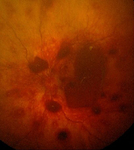
Retinal hemorrhages in abusive head trauma are usually widespread and multilayered, as seen in this image
From the personal collection of Alice Newton, MD; used with permission
See this image in context in the following section/s:
Abusive head trauma in infants and young children
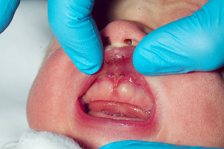
Torn labial frenulum with associated bruising in a neonate
Reproduced with permission from Gurung H et al. Labial frenum tear from instrumental delivery. Arch Dis Child. 2015 Aug;100(8):773
See this image in context in the following section/s:
Use of this content is subject to our disclaimer