Suspect hypothermia (core body temperature <95°F [<35°C]) based on the condition the patient is found in and/or the presence of risk factors.
Prehospital assessment
In a prehospital setting, use the four-stage original Swiss system to help estimate the patient’s core temperature at the scene (if this isn’t already available).[19]Paal P, Pasquier M, Darocha T, et al. Accidental hypothermia: 2021 update. Int J Environ Res Public Health. 2022;19(1):501.
https://www.mdpi.com/1660-4601/19/1/501
http://www.ncbi.nlm.nih.gov/pubmed/35010760?tool=bestpractice.com
Vital signs can be present even when core temperature is below 75°F (24°C).[44]Pasquier M, Zurron N, Weith B, et al. Deep accidental hypothermia with core temperature below 24°C presenting with vital signs. High Alt Med Biol. 2014 Apr;15(1):58-63.
http://www.ncbi.nlm.nih.gov/pubmed/24527793?tool=bestpractice.com
[45]Panchal AR, Bartos JA, Cabañas JG, et al. Part 3: adult basic and advanced life support: 2020 American Heart Association guidelines for cardiopulmonary resuscitation and emergency cardiovascular care. Circulation. 2020 Oct 20;142(16 Suppl 2):S366-468.
https://www.ahajournals.org/doi/full/10.1161/CIR.0000000000000916
http://www.ncbi.nlm.nih.gov/pubmed/33081529?tool=bestpractice.com
Stages of hypothermia are based on clinical signs (shivering, vital signs, level of consciousness) that roughly correlate to the patient’s core temperature and are used to guide management.[46]Durrer B, Brugger H, Syme D, et al. The medical on-site treatment of hypothermia: ICAR-MEDCOM recommendation. High Alt Med Biol. 2003 Spring;4(1):99-103.
http://www.ncbi.nlm.nih.gov/pubmed/12713717?tool=bestpractice.com
However, it is important to note that factors such as trauma, central nervous system failure, and substance misuse and overdose may impair shivering and consciousness, independent of a patient’s core temperature.[19]Paal P, Pasquier M, Darocha T, et al. Accidental hypothermia: 2021 update. Int J Environ Res Public Health. 2022;19(1):501.
https://www.mdpi.com/1660-4601/19/1/501
http://www.ncbi.nlm.nih.gov/pubmed/35010760?tool=bestpractice.com
Consider the revised Swiss system, which may simplify clinical staging in the field, if appropriate; the revised system incorporates risk of cardiac arrest as part of staging.[47]Musi ME, Sheets A, Zafren K, et al. Clinical staging of accidental hypothermia: The revised Swiss system: Recommendation of the International Commission for Mountain Emergency Medicine (ICAR MedCom). Resuscitation. 2021 May;162:182-7.
https://www.resuscitationjournal.com/article/S0300-9572(21)00096-4/fulltext
http://www.ncbi.nlm.nih.gov/pubmed/33675869?tool=bestpractice.com
See Cardiac arrest.
If the patient has been immersed in water, see Drowning.
History
Ask the prehospital team about the condition in which the patient was found. This may provide important clues to the diagnosis; for example, patients who are inappropriately dressed for a cold climate and have spent a long time outdoors or in a cold environment may be hypothermic.
Consider risk factors for hypothermia. These include:[2]van Veelen MJ, Brodmann Maeder M. Hypothermia in trauma. Int J Environ Res Public Health. 2021 Aug 18;18(16):8719.
https://www.mdpi.com/1660-4601/18/16/8719
http://www.ncbi.nlm.nih.gov/pubmed/34444466?tool=bestpractice.com
[16]Zhang P, Wiens K, Wang R, et al. Cold weather conditions and risk of hypothermia among people experiencing homelessness: implications for prevention strategies. Int J Environ Res Public Health. 2019 Sep 5;16(18):3259.
https://www.mdpi.com/1660-4601/16/18/3259
http://www.ncbi.nlm.nih.gov/pubmed/31491874?tool=bestpractice.com
[19]Paal P, Pasquier M, Darocha T, et al. Accidental hypothermia: 2021 update. Int J Environ Res Public Health. 2022;19(1):501.
https://www.mdpi.com/1660-4601/19/1/501
http://www.ncbi.nlm.nih.gov/pubmed/35010760?tool=bestpractice.com
[27]Giesbrecht GG. Cold stress, near drowning and accidental hypothermia: a review. Aviat Space Environ Med. 2000 Jul;71(7):733-52.
http://www.ncbi.nlm.nih.gov/pubmed/10902937?tool=bestpractice.com
[31]Leslie K, Sessler DI. Perioperative hypothermia in the high-risk surgical patient. Best Pract Res Clin Anaesthesiol. 2003 Dec;17(4):485-98.
http://www.ncbi.nlm.nih.gov/pubmed/14661653?tool=bestpractice.com
[34]Bridwell RE, Willis GC, Gottlieb M, et al. Decompensated hypothyroidism: a review for the emergency clinician. Am J Emerg Med. 2021 Jan;39:207-12.
http://www.ncbi.nlm.nih.gov/pubmed/33039222?tool=bestpractice.com
[35]Snijders BMG, Roos MJ, Keijsers CJPW. Incidences of underlying causes of hypothermia in older patients in the emergency department: a systematic review. Eur Geriatr Med. 2023 Jun;14(3):411-20.
https://pmc.ncbi.nlm.nih.gov/articles/PMC10261225
http://www.ncbi.nlm.nih.gov/pubmed/37191873?tool=bestpractice.com
[36]Singer D. Pediatric hypothermia: an ambiguous issue. Int J Environ Res Public Health. 2021 Oct 31;18(21):11484.
https://www.mdpi.com/1660-4601/18/21/11484
http://www.ncbi.nlm.nih.gov/pubmed/34769999?tool=bestpractice.com
[37]Bright F, Gilbert JD, Winskog C, et al. Additional risk factors for lethal hypothermia. J Forensic Leg Med. 2013 Aug;20(6):595-7.
http://www.ncbi.nlm.nih.gov/pubmed/23910840?tool=bestpractice.com
[38]Zonnenberg C, Bueno-de-Mesquita JM, Ramlal D, et al. Hypothermia due to antipsychotic medication: a systematic review. Front Psychiatry. 2017;8:165.
https://www.frontiersin.org/journals/psychiatry/articles/10.3389/fpsyt.2017.00165/full
http://www.ncbi.nlm.nih.gov/pubmed/28936184?tool=bestpractice.com
Physical exam including core temperature measurement
Examine and move the patient very carefully while you are assessing them. Keep the patient in a supine position if they have features of moderate or severe hypothermia (e.g., they have stopped shivering or have a reduced level of consciousness) as movement can precipitate ventricular fibrillation, especially if the patient’s temperature is <82°F (<28°C).[20]Lott C, Truhlář A, Alfonzo A, et al. European Resuscitation Council guidelines 2021: cardiac arrest in special circumstances. Resuscitation. 2021 Apr;161:152-219.
https://www.resuscitationjournal.com/article/S0300-9572(21)00064-2/fulltext
http://www.ncbi.nlm.nih.gov/pubmed/33773826?tool=bestpractice.com
[48]Dow J, Giesbrecht GG, Danzl DF, et al. Wilderness Medical Society clinical practice guidelines for the out-of-hospital evaluation and treatment of accidental hypothermia: 2019 update. Wilderness Environ Med. 2019 Dec;30(4 Suppl):S47-69.
https://www.wemjournal.org/article/S1080-6032(19)30173-5/fulltext
http://www.ncbi.nlm.nih.gov/pubmed/31740369?tool=bestpractice.com
Check for vital signs (including a carotid pulse) for up to 1 minute.[20]Lott C, Truhlář A, Alfonzo A, et al. European Resuscitation Council guidelines 2021: cardiac arrest in special circumstances. Resuscitation. 2021 Apr;161:152-219.
https://www.resuscitationjournal.com/article/S0300-9572(21)00064-2/fulltext
http://www.ncbi.nlm.nih.gov/pubmed/33773826?tool=bestpractice.com
[48]Dow J, Giesbrecht GG, Danzl DF, et al. Wilderness Medical Society clinical practice guidelines for the out-of-hospital evaluation and treatment of accidental hypothermia: 2019 update. Wilderness Environ Med. 2019 Dec;30(4 Suppl):S47-69.
https://www.wemjournal.org/article/S1080-6032(19)30173-5/fulltext
http://www.ncbi.nlm.nih.gov/pubmed/31740369?tool=bestpractice.com
Measure and monitor vital signs as part of ongoing assessment, including: blood pressure; pulse rate; respiratory rate; and oxygen saturations. Be aware that vital signs may be very difficult to detect in a patient with hypothermia, especially in the prehospital setting; a very hypothermic patient may appear dead but still survive with resuscitation.[20]Lott C, Truhlář A, Alfonzo A, et al. European Resuscitation Council guidelines 2021: cardiac arrest in special circumstances. Resuscitation. 2021 Apr;161:152-219.
https://www.resuscitationjournal.com/article/S0300-9572(21)00064-2/fulltext
http://www.ncbi.nlm.nih.gov/pubmed/33773826?tool=bestpractice.com
[45]Panchal AR, Bartos JA, Cabañas JG, et al. Part 3: adult basic and advanced life support: 2020 American Heart Association guidelines for cardiopulmonary resuscitation and emergency cardiovascular care. Circulation. 2020 Oct 20;142(16 Suppl 2):S366-468.
https://www.ahajournals.org/doi/full/10.1161/CIR.0000000000000916
http://www.ncbi.nlm.nih.gov/pubmed/33081529?tool=bestpractice.com
[48]Dow J, Giesbrecht GG, Danzl DF, et al. Wilderness Medical Society clinical practice guidelines for the out-of-hospital evaluation and treatment of accidental hypothermia: 2019 update. Wilderness Environ Med. 2019 Dec;30(4 Suppl):S47-69.
https://www.wemjournal.org/article/S1080-6032(19)30173-5/fulltext
http://www.ncbi.nlm.nih.gov/pubmed/31740369?tool=bestpractice.com
[49]Foggle JL. Accidental hypothermia: 'You're not dead until you're warm and dead'. R I Med J (2013). 2019 Feb 1;102(1):28-32.
http://rimed.org/rimedicaljournal/2019/02/2019-02-28-wilderness-foggle.pdf
http://www.ncbi.nlm.nih.gov/pubmed/30709071?tool=bestpractice.com
Other causes of cardiac arrest may need to be excluded; cardiac arrest is unlikely to be solely due to hypothermia unless the core temperature is less than 82°F (28°C).[19]Paal P, Pasquier M, Darocha T, et al. Accidental hypothermia: 2021 update. Int J Environ Res Public Health. 2022;19(1):501.
https://www.mdpi.com/1660-4601/19/1/501
http://www.ncbi.nlm.nih.gov/pubmed/35010760?tool=bestpractice.com
Patients often show signs of confusion or impaired judgment. Additionally, they may be shivering, have increased urinary frequency, and show signs of frostbite on their skin. See Frostbite.
Note that shivering will be absent once the patient’s core temperature drops below a certain level; the threshold varies between patients but is typically 82°F to 90°F (28°C to 32°C).[48]Dow J, Giesbrecht GG, Danzl DF, et al. Wilderness Medical Society clinical practice guidelines for the out-of-hospital evaluation and treatment of accidental hypothermia: 2019 update. Wilderness Environ Med. 2019 Dec;30(4 Suppl):S47-69.
https://www.wemjournal.org/article/S1080-6032(19)30173-5/fulltext
http://www.ncbi.nlm.nih.gov/pubmed/31740369?tool=bestpractice.com
However, it is important to be aware that factors such as trauma, central nervous system failure, and substance misuse and overdose may impair shivering and consciousness, independent of a patient’s core temperature.
Look for any signs of the underlying cause of hypothermia. For example:[45]Panchal AR, Bartos JA, Cabañas JG, et al. Part 3: adult basic and advanced life support: 2020 American Heart Association guidelines for cardiopulmonary resuscitation and emergency cardiovascular care. Circulation. 2020 Oct 20;142(16 Suppl 2):S366-468.
https://www.ahajournals.org/doi/full/10.1161/CIR.0000000000000916
http://www.ncbi.nlm.nih.gov/pubmed/33081529?tool=bestpractice.com
Self-harm. Consider this particularly if the patient has a reduced level of consciousness or been immersed in water. Check for any signs of drug overdose or alcohol intoxication. See Overview of substance use disorders and overdose.
Acute illness (e.g., stroke) or injury that has resulted in the patient lying on the ground outdoors for a long period of time.
Clinical signs correlate approximately to the patient’s core temperature.[48]Dow J, Giesbrecht GG, Danzl DF, et al. Wilderness Medical Society clinical practice guidelines for the out-of-hospital evaluation and treatment of accidental hypothermia: 2019 update. Wilderness Environ Med. 2019 Dec;30(4 Suppl):S47-69.
https://www.wemjournal.org/article/S1080-6032(19)30173-5/fulltext
http://www.ncbi.nlm.nih.gov/pubmed/31740369?tool=bestpractice.com
However, an individual patient’s response to hypothermia may vary considerably; clinical signs can only provide an estimate of core temperature. Urgent critical care support is required for any patient with severe hypothermia.
Core temperature
Do not use a standard clinical thermometer to measure core temperature. This may be inadequate as it will not measure temperatures below 94°F (34.4°C). Conventional mercury thermometers are also not recommended, owing to the risk of breakage and poisoning.
Where feasible (usually in hospital) the 2019 Wilderness Medical Society guidelines and the 2021 European Resuscitation Council guidelines recommend:
Preferably: an esophageal probe.[20]Lott C, Truhlář A, Alfonzo A, et al. European Resuscitation Council guidelines 2021: cardiac arrest in special circumstances. Resuscitation. 2021 Apr;161:152-219.
https://www.resuscitationjournal.com/article/S0300-9572(21)00064-2/fulltext
http://www.ncbi.nlm.nih.gov/pubmed/33773826?tool=bestpractice.com
[48]Dow J, Giesbrecht GG, Danzl DF, et al. Wilderness Medical Society clinical practice guidelines for the out-of-hospital evaluation and treatment of accidental hypothermia: 2019 update. Wilderness Environ Med. 2019 Dec;30(4 Suppl):S47-69.
https://www.wemjournal.org/article/S1080-6032(19)30173-5/fulltext
http://www.ncbi.nlm.nih.gov/pubmed/31740369?tool=bestpractice.com
An esophageal probe correlates well with the temperature of the pulmonary artery and is the preferred method when available.[19]Paal P, Pasquier M, Darocha T, et al. Accidental hypothermia: 2021 update. Int J Environ Res Public Health. 2022;19(1):501.
https://www.mdpi.com/1660-4601/19/1/501
http://www.ncbi.nlm.nih.gov/pubmed/35010760?tool=bestpractice.com
[48]Dow J, Giesbrecht GG, Danzl DF, et al. Wilderness Medical Society clinical practice guidelines for the out-of-hospital evaluation and treatment of accidental hypothermia: 2019 update. Wilderness Environ Med. 2019 Dec;30(4 Suppl):S47-69.
https://www.wemjournal.org/article/S1080-6032(19)30173-5/fulltext
http://www.ncbi.nlm.nih.gov/pubmed/31740369?tool=bestpractice.com
This is usually only possible in critically ill patients as readings must be obtained from the lower third of the esophagus when the airway is secured (i.e., tracheal tube or a supraglottic device with an esophageal channel in place).
Alternatively: a low-reading tympanic membrane thermistor-based thermometer (where the thermistor touches the tympanic membrane) if the patient is spontaneously breathing.[20]Lott C, Truhlář A, Alfonzo A, et al. European Resuscitation Council guidelines 2021: cardiac arrest in special circumstances. Resuscitation. 2021 Apr;161:152-219.
https://www.resuscitationjournal.com/article/S0300-9572(21)00064-2/fulltext
http://www.ncbi.nlm.nih.gov/pubmed/33773826?tool=bestpractice.com
[48]Dow J, Giesbrecht GG, Danzl DF, et al. Wilderness Medical Society clinical practice guidelines for the out-of-hospital evaluation and treatment of accidental hypothermia: 2019 update. Wilderness Environ Med. 2019 Dec;30(4 Suppl):S47-69.
https://www.wemjournal.org/article/S1080-6032(19)30173-5/fulltext
http://www.ncbi.nlm.nih.gov/pubmed/31740369?tool=bestpractice.com
Bladder catheter temperature sensors can be used in patients who require a urinary catheter, but bladder and rectal temperature lag behind core temperature and are only recommended for stable patients in a hospital setting.[19]Paal P, Pasquier M, Darocha T, et al. Accidental hypothermia: 2021 update. Int J Environ Res Public Health. 2022;19(1):501.
https://www.mdpi.com/1660-4601/19/1/501
http://www.ncbi.nlm.nih.gov/pubmed/35010760?tool=bestpractice.com
[20]Lott C, Truhlář A, Alfonzo A, et al. European Resuscitation Council guidelines 2021: cardiac arrest in special circumstances. Resuscitation. 2021 Apr;161:152-219.
https://www.resuscitationjournal.com/article/S0300-9572(21)00064-2/fulltext
http://www.ncbi.nlm.nih.gov/pubmed/33773826?tool=bestpractice.com
Never measure rectal temperature if the patient is in a cold environment. This method requires the patient to be further exposed, which will increase heat loss and potentially worsen hypothermia.
Investigations
Investigations in the workup of accidental hypothermia are not diagnostic, but do help to guide acute management.
ECG
Continuous ECG monitoring is essential for detecting arrhythmias, which may be fatal. Where possible, ECG monitoring should also be used to detect cardiac arrest.[20]Lott C, Truhlář A, Alfonzo A, et al. European Resuscitation Council guidelines 2021: cardiac arrest in special circumstances. Resuscitation. 2021 Apr;161:152-219.
https://www.resuscitationjournal.com/article/S0300-9572(21)00064-2/fulltext
http://www.ncbi.nlm.nih.gov/pubmed/33773826?tool=bestpractice.com
[48]Dow J, Giesbrecht GG, Danzl DF, et al. Wilderness Medical Society clinical practice guidelines for the out-of-hospital evaluation and treatment of accidental hypothermia: 2019 update. Wilderness Environ Med. 2019 Dec;30(4 Suppl):S47-69.
https://www.wemjournal.org/article/S1080-6032(19)30173-5/fulltext
http://www.ncbi.nlm.nih.gov/pubmed/31740369?tool=bestpractice.com
Arrhythmias can occur at any stage of hypothermia, and also during rewarming. Initially, in mild hypothermia, the ECG may show tachycardia. In more severe cases of hypothermia, the ECG may show progressive sinus bradycardia, atrial or ventricular fibrillation, junctional rhythms, ST segment changes, T-wave inversion, prolongation of the QT interval, and eventually asystole.[11]Mallet ML. Pathophysiology of accidental hypothermia. QJM. 2002 Dec;95(12):775-85.
https://academic.oup.com/qjmed/article/95/12/775/1572021
http://www.ncbi.nlm.nih.gov/pubmed/12454320?tool=bestpractice.com
With the exception of ventricular fibrillation, these changes are likely to improve without treatment as the patient’s core temperature increases.[20]Lott C, Truhlář A, Alfonzo A, et al. European Resuscitation Council guidelines 2021: cardiac arrest in special circumstances. Resuscitation. 2021 Apr;161:152-219.
https://www.resuscitationjournal.com/article/S0300-9572(21)00064-2/fulltext
http://www.ncbi.nlm.nih.gov/pubmed/33773826?tool=bestpractice.com
[48]Dow J, Giesbrecht GG, Danzl DF, et al. Wilderness Medical Society clinical practice guidelines for the out-of-hospital evaluation and treatment of accidental hypothermia: 2019 update. Wilderness Environ Med. 2019 Dec;30(4 Suppl):S47-69.
https://www.wemjournal.org/article/S1080-6032(19)30173-5/fulltext
http://www.ncbi.nlm.nih.gov/pubmed/31740369?tool=bestpractice.com
J waves (or Osborn waves) occur in most, but not all, patients.[50]Higuchi S, Takahashi T, Kabeya Y, et al. J waves in accidental hypothermia. Circ J. 2014;78(1):128-34.
https://www.jstage.jst.go.jp/article/circj/78/1/78_CJ-13-0704/_article
http://www.ncbi.nlm.nih.gov/pubmed/24200873?tool=bestpractice.com
However, they do not correlate well with temperature.[51]Urdang M, Shoenberger JM, Triamarit P, et al. ECG analysis in accidental urban hypothermia. J Emerg Med. 2009 Aug;37(2):235.
[Figure caption and citation for the preceding image starts]: A 12-lead ECG obtained from a hypothermic patient; note, Osborn waves (arrows), which have an extra deflection at the end of the QRS complexAydin M, Gursurer M, Bayraktaroglu T, et al. Tex Heart Inst J. 2005;32(1):105 [Citation ends].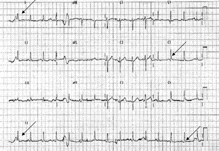
Laboratory tests
Initial investigations should include: arterial blood gas (ABG), blood glucose, and blood chemistries. Further tests are less useful for the acute assessment and management of hypothermia but generally include a complete blood count (CBC) and clotting screen.
An ABG may show respiratory alkalosis, metabolic acidosis, or a mixed picture. As core temperature decreases, respiration is depressed, resulting in hypoxemia and hypercapnia. A combined respiratory and metabolic acidosis occurs as a result of hypoventilation, retention of carbon dioxide, decreased bicarbonate, impaired hepatic metabolism of organic acid production (owing to impaired hepatic perfusion), and increased lactic acid production. It is important to note that blood pH rises by 0.015 for every 1.8°F (1°C) drop in body temperature. In general, use blood gas results without adjustment for temperature to guide treatment decisions.[52]Darocha T, Kosiński S, Jarosz A, et al. Should capnography be used as a guide for choosing a ventilation strategy in circulatory shock caused by severe hypothermia? Observational case-series study. Scand J Trauma Resusc Emerg Med. 2017 Feb 15;25(1):15.
https://sjtrem.biomedcentral.com/articles/10.1186/s13049-017-0357-1
http://www.ncbi.nlm.nih.gov/pubmed/28202085?tool=bestpractice.com
Glucose levels may be normal, high (owing to increased secretion of stress hormones - cortisol, growth hormones, and catecholamines - and reduced insulin secretion, together with increased peripheral resistance to insulin), or low (owing to cold-induced inhibition of hepatic glucose production). Monitor blood glucose even after the patient is normoglycemic because rebound hypoglycemia may develop when normal insulin production resumes. Treat hypoglycemia promptly. Hypoglycemia can stop shivering (because the central control of shivering is dependent on glucose), leading to subsequent heat loss.[53]Gale EA, Bennett T, Green JH, et al. Hypoglycaemia, hypothermia and shivering in man. Clin Sci (Lond). 1981 Oct;61(4):463-9.
http://www.ncbi.nlm.nih.gov/pubmed/7026128?tool=bestpractice.com
Renal function may be impaired due to dehydration, cold exposure, or rhabdomyolysis.
Hypokalemia may occur as a result of hypothermia or the associated treatment. Hyperkalemia may occur during rewarming. For a patient in cardiac arrest, hyperkalemia can also indicate that hypoxia preceded hypothermia (e.g., if the patient was found in an avalanche).[48]Dow J, Giesbrecht GG, Danzl DF, et al. Wilderness Medical Society clinical practice guidelines for the out-of-hospital evaluation and treatment of accidental hypothermia: 2019 update. Wilderness Environ Med. 2019 Dec;30(4 Suppl):S47-69.
https://www.wemjournal.org/article/S1080-6032(19)30173-5/fulltext
http://www.ncbi.nlm.nih.gov/pubmed/31740369?tool=bestpractice.com
Initial serum potassium >12 mEq/L (>12 mmol/L) is associated with irreversible death if the patient is in cardiac arrest.[48]Dow J, Giesbrecht GG, Danzl DF, et al. Wilderness Medical Society clinical practice guidelines for the out-of-hospital evaluation and treatment of accidental hypothermia: 2019 update. Wilderness Environ Med. 2019 Dec;30(4 Suppl):S47-69.
https://www.wemjournal.org/article/S1080-6032(19)30173-5/fulltext
http://www.ncbi.nlm.nih.gov/pubmed/31740369?tool=bestpractice.com
Serum potassium is part of the HOPE (Hypothermia Outcome Prediction after ECLS rewarming for hypothermic arrested patients) score for prognostication of successful rewarming.[20]Lott C, Truhlář A, Alfonzo A, et al. European Resuscitation Council guidelines 2021: cardiac arrest in special circumstances. Resuscitation. 2021 Apr;161:152-219.
https://www.resuscitationjournal.com/article/S0300-9572(21)00064-2/fulltext
http://www.ncbi.nlm.nih.gov/pubmed/33773826?tool=bestpractice.com
[48]Dow J, Giesbrecht GG, Danzl DF, et al. Wilderness Medical Society clinical practice guidelines for the out-of-hospital evaluation and treatment of accidental hypothermia: 2019 update. Wilderness Environ Med. 2019 Dec;30(4 Suppl):S47-69.
https://www.wemjournal.org/article/S1080-6032(19)30173-5/fulltext
http://www.ncbi.nlm.nih.gov/pubmed/31740369?tool=bestpractice.com
[
Hypothermia outcome prediction after ECLS (HOPE) score
Opens in new window
]
A CBC may show elevated hemoglobin and hematocrit, and low platelet and white blood cell counts.
Prothrombin time and partial thromboplastin time (PTT) tend to be prolonged, although the cause for this is unknown.[54]Breen EG, Coghlan JG, Egan E, et al. Impaired coagulation in accidental hypothermia of the elderly. Age Ageing. 1988 Sep;17(5):343-6.
http://www.ncbi.nlm.nih.gov/pubmed/3232589?tool=bestpractice.com
Imaging
A chest x-ray is particularly important if patients have an altered level of consciousness. It may show pulmonary edema or infiltrates.
If the patient has been immersed in water, it may show inhaled foreign bodies, such as false teeth or debris from the water, which will need to be removed. See Foreign body aspiration.
Investigations to consider
Serum creatinine kinase and myoglobin levels should be checked for rhabdomyolysis if the patient may have been lying on the ground outdoors for a long time and they have not been immersed in water. See Rhabdomyolysis.
If vital signs are undetectable, use ultrasound and end-tidal CO₂, where possible, to confirm cardiac arrest.[20]Lott C, Truhlář A, Alfonzo A, et al. European Resuscitation Council guidelines 2021: cardiac arrest in special circumstances. Resuscitation. 2021 Apr;161:152-219.
https://www.resuscitationjournal.com/article/S0300-9572(21)00064-2/fulltext
http://www.ncbi.nlm.nih.gov/pubmed/33773826?tool=bestpractice.com
[48]Dow J, Giesbrecht GG, Danzl DF, et al. Wilderness Medical Society clinical practice guidelines for the out-of-hospital evaluation and treatment of accidental hypothermia: 2019 update. Wilderness Environ Med. 2019 Dec;30(4 Suppl):S47-69.
https://www.wemjournal.org/article/S1080-6032(19)30173-5/fulltext
http://www.ncbi.nlm.nih.gov/pubmed/31740369?tool=bestpractice.com