Images and videos
Images


Evaluation of pleuritis
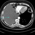
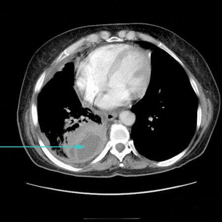
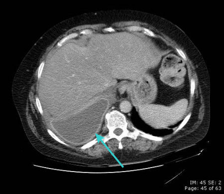
CT scan showing large right pleural effusion
From the collection of Dr Nicholas Maskell; used with permission
See this image in context in the following section/s:
Evaluation of pleuritis
CT scan showing loculated pleural effusion
From the collection of Dr Ami Rubinowitz; used with permission
See this image in context in the following section/s:
Evaluation of pleuritis
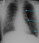
Large left pneumothorax with visible pleural line and absence of lung markings beyond the line
From the collection of Dr Ami Rubinowitz; used with permission
See this image in context in the following section/s:
Evaluation of pleuritis
Modified Wells criteria (score ≤4: PE unlikely; score >4: PE likely)
From Wells PS, Anderson DR, Rodger M, et al. Thromb Haemost. 2000;83:416. Used with permission
See this image in context in the following section/s:
Evaluation of pleuritis
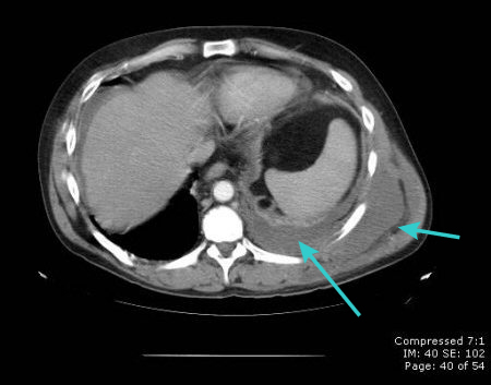
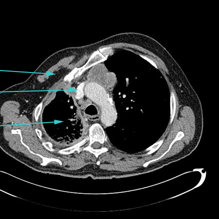
CT scan of chest showing empyema necessitans (long arrow), a chronic untreated empyema that has eroded through the thoracic cage and formed a subcutaneous abscess (short arrow)
From the collection of Dr Ami Rubinowitz; used with permission
See this image in context in the following section/s:

Evaluation of pleuritis
CT scan showing metastatic malignancy of pleura
From the collection of Dr Ami Rubinowitz; used with permission
See this image in context in the following section/s:
Evaluation of pleuritis
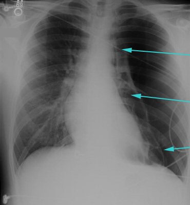
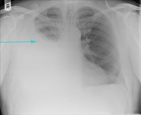
CXR showing large right pleural effusion
From the collection of Dr Kathryn Bateman; used with permission
See this image in context in the following section/s:

Evaluation of pleuritis
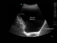
Thoracic ultrasound image of large, simple pleural effusion
From the collection of Dr Nicholas Maskell; used with permission
See this image in context in the following section/s:
Evaluation of pleuritis
Schematic showing a summary of the diagnostic approach (CXR=chest x-ray, PE-pulmonary embolism, EKG=electrocardiogram)
Courtesy of Dr Ami Rubinowitz; used with permission. Updated November 2023 by BMJ Best Practice Editorial Team
See this image in context in the following section/s:

Evaluation of pleuritis
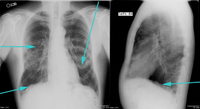
PA and lateral CXR showing calcified pleural plaques
From the collection of Dr Ami Rubinowitz; used with permission
See this image in context in the following section/s:
Evaluation of pleuritis
CT scan showing empyema with split pleura sign (enhancement of the thickened inner visceral and outer parietal pleura separated by a collection of pleural fluid)
From the collection of Dr Ami Rubinowitz; used with permission
See this image in context in the following section/s:
Evaluation of pleuritis
CT scan showing circumferential right-sided pleural thickening with significant volume loss due to mesothelioma
From the collection of Dr Nicholas Maskell; used with permission
See this image in context in the following section/s:
Videos
 Needle decompression of tension pneumothorax: animated demonstration
How to decompress a tension pneumothorax. Demonstrates insertion of a large-bore intravenous catheter into the fourth intercostal space in an adult.
 Insertion of intercostal drain, open technique: animated demonstration
How to insert an intercostal (chest) drain using the open technique. Video demonstrates: tube selection, how to identify the site for drain insertion, how to make the correct incision, how to insert the intercostal drain, how to secure the drain, and postprocedure care.
 Insertion of intercostal drain, Seldinger technique: animated demonstration
How to insert an intercostal (chest) drain using the Seldinger technique. Video demonstrates: how to identify a safe site for insertion; use of an introducer needle, guidewire, dilators, and intercostal drain; how to confirm drain position; and postprocedure care.
Use of this content is subject to our disclaimer