Etiology
The pleura and the pleural space are affected by a variety of systemic disease processes as well as local diseases affecting the lung.[3] Some rarer causes arise from the pleural space itself.
Pulmonary
Two-thirds of pleural infections arise from underlying lung infections or penetrating thoracic trauma. Other sources of pleural infections are vascular dissemination or extension from an intra-abdominal source.[4]
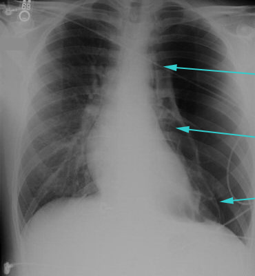
Pneumothorax: when the pleural space is disrupted air collects between the parietal and visceral pleura. This is seen as a pleural line beyond which no parenchymal markings are visible on a chest radiograph. When this occurs in the setting of accompanying pleural fluid (a hydropneumothorax), the fluid exposed to air will often have increased eosinophils as a result.
Viral: coxsackie B virus is a common cause of infectious pleuritis.[3] Echovirus can cause the syndrome Bornholm pleurodynia, manifesting as pleuritis, fever, and chest muscle spasms; the condition occurs in the late summer and affects adolescents and young adults. Other viral etiologies are influenza, parainfluenzae, respiratory syncytial virus, COVID-19, and cytomegalovirus.
Bacterial: Streptococcus species, Staphylococcus aureus, enteric gram-negative bacilli, and anaerobes are the principal organisms causing pleural infections. They invariably result in the formation of empyema if not promptly drained.[5]
Pulmonary embolism: when an effusion develops because of nearby lung infarction, the fluid is typically sanguineous or serosanguineous and may have predominance of neutrophils or eosinophils.[6][7]
Asbestos-related benign pleural disease: asbestos exposure can result in parietal pleural plaques or diffuse pleural thickening, usually many (>18) years after exposure. Benign asbestos pleural effusions, however, can occur as early as 1 year after exposure and are exudative with elevated eosinophils.[8] These usually self remit but may do so slowly over many months. Initial presentation is usually with dyspnea on exertion. Rarely patients can present with pleuritic chest pain.[9][Figure caption and citation for the preceding image starts]: Large left pneumothorax with visible pleural line and absence of lung markings beyond the lineFrom the collection of Dr Ami Rubinowitz; used with permission [Citation ends].
Systemic
Infection: tuberculous pleuritis is the most common form of extrapulmonary tuberculosis (TB) infection.[10] It is uncommon in the US, but is increasingly prevalent worldwide.[11] A definitive diagnosis of TB pleuritis can only be confirmed by identification of Mycobacterium tuberculosis by microscopy; culture of sputum, pleural fluid, or pleural biopsy specimens; or nucleic acid amplification tests (NAATs).[12][13][14] Unfortunately, acid-fast smears and culture of sputum samples have poor sensitivity in patients with pleural TB.[15][16] NAATs should be performed on at least one respiratory specimen, or specimen collected from sites of suspected extrapulmonary TB, when a diagnosis of TB is being considered.[14][17] The pleural fluid biomarker, adenosine deaminase, has a high sensitivity and specificity for the identification of pleural TB (92% and 90%, respectively), and could facilitate prompt initiation of treatment in areas of high TB prevalence.[12]
Malignancy: primary lung cancer, mesothelioma (as a result of asbestos exposure), or metastasis from lung, breast, lymphoma, gastrointestinal, or genitourinary tumors.[18]
Connective tissue disorders: a major cause of autoimmune pleuritis. The most common connective tissue disorders are systemic lupus erythematosus, rheumatoid arthritis, and Sjogren syndrome. In one retrospective review, 8.5% of patients with nonspecific pleuritis on thoracoscopic biopsy were diagnosed with Sjogren syndrome.[19]
Drug reactions: drugs such as hydralazine, procainamide, isoniazid, methyldopa, or chlorpromazine are associated with development of lupus pleuritis. Other drugs such as minoxidil, beta blockers, amiodarone, bleomycin, methysergide, methotrexate, cyclophosphamide, valproic acid, or nitrofurantoin also cause pleuritis through unclear mechanisms.[20] Pneumotox: drug-induced respiratory diseases Opens in new window
Other conditions that may cause pleuritis, although it is rarely the primary presenting symptom, include uremia, familial Mediterranean fever, and other intra-abdominal processes such as cirrhosis or pancreatitis.[21][22][23]
Cardiovascular
Postcardiac injury syndrome: an autoimmune inflammatory process involving pleura and pericardium secondary to cardiac injury (either acute coronary syndrome or cardiac surgery/trauma).[24]
Aortic dissection: rare cause of pleuritis and is due to blood leaking into the pleural space.
Use of this content is subject to our disclaimer