Images and videos
Images


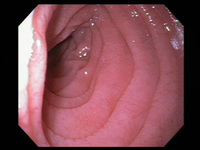

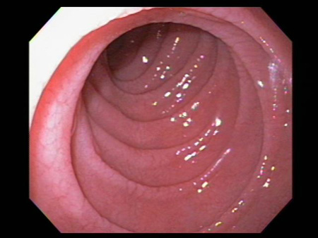
Celiac disease
Scalloping of the duodenal mucosa in a patient with celiac disease
From the personal collection of DA Leffler; used with permission
See this image in context in the following section/s:
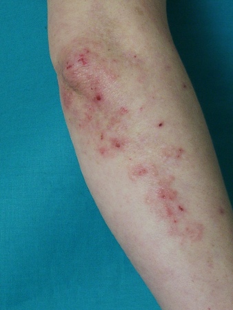
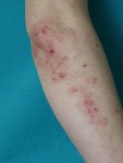
Celiac disease
Dermatitis herpetiformis: typical lesions on extensor surface of forearm
From the collection of Adam Reich MD, PhD
See this image in context in the following section/s:

Celiac disease
Scalloping of the duodenal mucosa in a patient with celiac disease
From the personal collection of DA Leffler; used with permission
See this image in context in the following section/s:

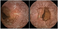
Celiac disease
Capsule endoscopy pictures of ulcerative jejunitis in a patient with celiac disease
From the personal collection of Amelie Therrien; used with permission
See this image in context in the following section/s:
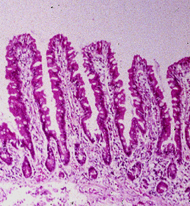
Celiac disease
Histologic image of small intestinal villi showing resolution of intestinal injury on gluten-free diet
From the personal collection of DA Leffler; used with permission
See this image in context in the following section/s:

Celiac disease
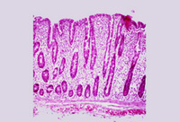
Histologic image of small intestinal villous atrophy and crypt hyperplasia
From the personal collection of DA Leffler; used with permission
See this image in context in the following section/s:
Celiac disease
Photograph of small intestinal villi affected by celiac disease
From the personal collection of DA Leffler; used with permission
See this image in context in the following section/s:
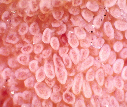
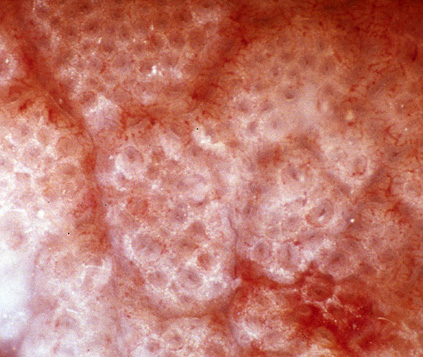
Celiac disease
Photograph of normal small intestinal villi
From the personal collection of DA Leffler; used with permission
See this image in context in the following section/s:
Use of this content is subject to our disclaimer