Approach
The diagnosis of keloid scarring is primarily clinical. A skin biopsy is rarely required and must be weighed against the risk of making the condition worse.
Clinical history
Unlike hypertrophic scarring, keloid is not typically associated with skin contractures.
Typical findings include:[19]
An inciting event (e.g., body piercing, surgery, or trauma in a predisposed individual)
Personal or family history of keloids
High-risk skin type
Symptoms of itch and pain
Gradual increase in size of lesions
Lack of spontaneous regression.
[Figure caption and citation for the preceding image starts]: Stable keloid due to ear piercingFrom the collection of Professor Andrew Burd, used with permission [Citation ends].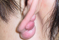
Physical exam
Site
Typically located over high tension bodily areas (e.g., upper back/deltoid, presternal, lower abdominal regions) and over bony prominences.
Earlobes are also a common location, especially after ear piercing.[19]
[Figure caption and citation for the preceding image starts]: A typical presternal keloid; presence of central bridging, an elevated rolled edge and flattened peripheral extensionsFrom the collection of Professor Andrew Burd, used with permission [Citation ends].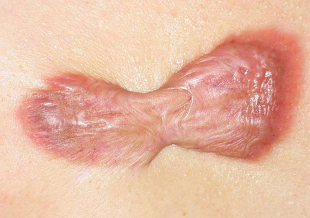 [Figure caption and citation for the preceding image starts]: Keloid reaction following a vaccination in the deltoid region; following surgical excision the entire length of the scar developed a keloid-like appearanceFrom the collection of Professor Andrew Burd, used with permission [Citation ends].
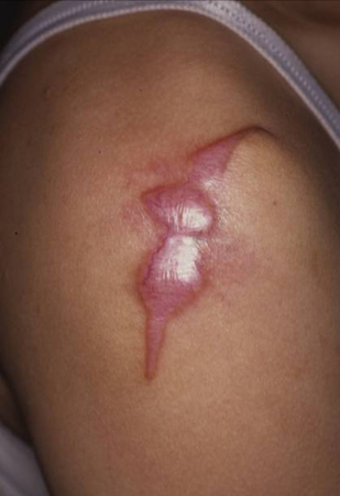
Texture or surface
The surface of the scar is typically smooth and shiny with an overhanging edge.
Consistency
Most keloid scars tend to be firm in consistency, especially if they are long-standing.
Color
Keloid scars may exhibit a range of colors depending on the normal skin pigmentation. During the active-growth phase, the scar may be erythematous, indicating increased blood flow. As the growth slows and begins to stabilize, the keloid may become more similar in color to the surrounding skin. [Figure caption and citation for the preceding image starts]: Red, elevated keloid; edges blend into the surrounding skin; arose in a vaccination site in the left upper armFrom the collection of Professor Andrew Burd, used with permission [Citation ends].

Investigations
Diagnosis is based on clinical findings.[17][19] The main reason to perform a skin biopsy is when there is doubt about the nature of the lesion or clinical suspicion of malignancy (e.g., rapid growth).[21]
The most important differential diagnosis is dermatofibrosarcoma, which typically involves more subdermal spread (the term subdermal is used rather than subcutaneous to underscore the pattern of growth). Clinically, the skin can appear to be normal on inspection but has a palpable tumor that arises from the dermis and spreads into the underlying tissue.
The need for histologic exam following keloidectomy should be discussed with individual patients. The risk of misdiagnosis of other pathology as keloid scar has been estimated to be 1.06%.[22]
Use of this content is subject to our disclaimer