Approach
The diagnosis of common warts is usually based on clinical findings. The clinical presentation of common warts is generally consistent. A common wart should be suspected in a patient with an enlarging:[1][2]
Rough papule on the hand, finger, periungual, or other skin area
Filiform papule on the skin of the body or face
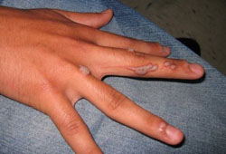
Flat pink, slightly scaly papule on the face or digits.[Figure caption and citation for the preceding image starts]: Verrucous papules on the fingers and periungual skin of the second, third, and fourth digits of a young adultCourtesy of Daniela Kroshinsky, MD, personal clinical files [Citation ends].
History and examination
Patients generally report a rough, scaly papule on the skin or around the nail fold or a stalk-like papule on the skin, enlarging over weeks to months. Occasionally, the rough papule may fissure, bleed, and become painful, especially when scratched or otherwise manipulated. Lesions may range in size from pinpoint to 1 cm, averaging 5 mm. Sometimes multiple, similar, smaller lesions develop following the appearance of the initial lesion.[1][2]
Examination reveals a single grayish-white or light-brown rough, hyperkeratotic papule on the fingers; around the nail fold; or on another skin area of the body. Sometimes lesions occur in clusters. Some lesions may be fissured or have a hemorrhagic crust after bleeding, or an erythematous base. Light paring with a scalpel reveals tiny black dots on the surface after the hyperkeratotic debris is removed.[1] In the case of filiform warts, examination reveals single or multiple filiform lesions that appear to emerge from the skin as a stalk with several sharp spikes.[Figure caption and citation for the preceding image starts]: Verrucous papules on the fingers and periungual skin of the second, third, and fourth digits of a young adultCourtesy of Daniela Kroshinsky, MD, personal clinical files [Citation ends].[Figure caption and citation for the preceding image starts]: Verrucous papule on the knee of a childCourtesy of Daniela Kroshinsky, MD, personal clinical files [Citation ends].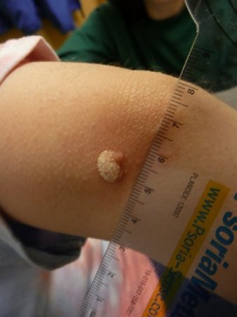
Investigations
In the overwhelming majority of infections occurring in healthy individuals, the history and physical examination alone are sufficient to make the diagnosis of common warts. The diagnosis is usually a clinical one. A helpful clue to the diagnosis is the demonstration of the tiny black dots on the surface.[1]
More procedures are required if the lesion is persistent or nonresponsive, because 70% to 80% of nonmelanoma skin cancers have been found to contain human papillomavirus. Biopsy is indicated if any of the following features are present:
Rapid growth
Nonhealing
Lack of treatment response
Bleeding
Crateriform and hyperkeratotic appearances.
These features apply especially when there is a history of immunosuppression, or chronic sun or radiation exposure, or when there is a previous history of actinic keratoses or skin malignancy. In immunocompromised patients, who tend to have many lesions, larger and more inflamed warts may need to be evaluated histopathologically. In rare cases, biopsy or culture for bacterial and/or fungal pathogens may be required, to rule out the rare verrucous presentations of deep fungal, bacterial, or mycobacterial infections.
Use of this content is subject to our disclaimer