Differentials
Common
Atopic dermatitis
History
recurrent dermatitis in areas of exposure to potential allergens such as skincare products or wool and lipid solvents (many patients have a history of milk allergy during infancy, and most also have other atopic disorders such as hay fever, atopic asthma, or a family history of atopic diseases)[50]
Exam
patients present with typical morphology and distribution of skin lesions: facial and extensor in infants and children, flexural lichenification in adolescents and adults; patients may present a wide range of other findings defined as minor criteria of atopic dermatitis[51]
1st investigation
- none:
diagnosis is clinical
Urticaria
History
sudden appearance of wheals with concomitant pruritus; patients can sometimes identify causative factors (e.g., cold, heat, water, food, drugs); detailed food diaries and in-depth drug reviews are typical starting points for diagnosis; list of possible factors is very long, so all patients should be thoroughly investigated
Exam
patients present typically with pruritic wheals; sometimes angioedema of lips, tongue, eyelids, or distal parts of extremities may be seen
1st investigation
- none:
diagnosis is clinical
Other investigations
- total serum IgE level:
elevated
More - allergen-specific IgE levels:
elevated
More - prick tests with specific allergens (aeroallergens, food allergens, medications):
wheals larger than control (histamine), usually diameter >5 mm
More - physical tests (exposure to pressure, cold, heat, UV light, physical activity, water):
induction of wheals after exposure to causative factor
More - exposure to drugs (e.g., acetylsalicylic acid):
induction of wheals after exposure to causative drug
More - stool microscopy:
detection of parasite eggs
More - antinuclear antibodies (ANA):
may be positive in autoimmune disease
More - test with autologous serum:
development of wheal after intradermal injection of the autologous serum
More - skin biopsy:
urticaria: edema of papillary dermis, sparse lymphocytic and eosinophilic perivascular and interstitial infiltrate may be present; urticarial vasculitis: signs of leukocytoclastic vasculitis
More
Insect bite
History
history of traveling, especially to wet regions
Exam
presence of dome-shaped urticarial papules, sometimes with a dot at summit of papule that represents site of the bite; lesions usually occur on unclothed areas of the skin; this is a short-lived reaction, but in some patients lesions may persist for weeks or months; similar symptoms are sometimes present in other family members
1st investigation
- none:
diagnosis is clinical
Other investigations
- hymenoptera allergy radioallergosorbent test (RAST):
positive in allergic urticaria (type I reaction)
Pinworm infection
History
perianal pruritus
Exam
erosions and secondary infections in perianal area
1st investigation
- adhesive tape test:
identification of eggs or adult worms on microscopy
Other investigations
Scabies
History
intense pruritus, especially at night in bed[57]
Exam
sites of predilection are the interdigital spaces, anterior axillary folds, nipples, umbilicus, elbows, and genitalia (especially scrotum); face and interscapular region are very rarely involved; the most diagnostic finding is an intact tunnel with a tiny dark dot (mite) at the end
1st investigation
- microscopy of skin scrapings:
presence of mites or their eggs
More
Other investigations
- dermoscopy:
identification of burrows; may see mite with characteristic "jet-plane" appearance and/or eggs
Lichen planus
History
nonspecific; sometimes a history of drug intake (e.g., gold salts) or hepatitis that can provoke lichen planus; frequently associated with intense pruritus
Exam
presence of small, smooth, violaceous, shiny, flat papules; lesions frequently involve inner aspects of wrists and back of the feet; Koebner phenomenon (lesions on areas or along lines of skin trauma) may be present; sometimes cutaneous lesions are associated with mucosal changes (lacy network on buccal mucosa or painful erosions); some patients may also have nail abnormalities manifesting mainly as longitudinal ridging
1st investigation
- skin biopsy:
band-like infiltrate of T cells at the epidermal-dermal junction, hyperplasia of granular layer in epidermis, and vacuolar changes in basal layer
More
Other investigations
- serologic test to detect antibodies against hepatitis B or C viruses:
detection of hepatitis B or C viral infection
More
Psoriasis
History
positive family history of psoriasis
Exam
primary lesion of psoriasis is a small, red, sharply demarcated papule covered with silvery scale; psoriatic papules may coalesce to form larger plaques; numerous clinical variants of psoriasis have been described; lesions are usually found on scalp, extensor areas (elbows, knees), and in sacroiliac area; nails may be involved (pitting, onycholysis, "oil spots")
1st investigation
- none:
diagnosis is clinical
Other investigations
- skin biopsy:
parakeratosis, epidermal hyperplasia, infiltration of epidermis with neutrophils (Munro microabscesses), elongated rete ridges
More
Skin xerosis
History
no specific features of skin dryness; diagnosis is based on skin appearance; usually older patients; may also be a nonspecific symptom of different skin or systemic diseases (e.g., atopic dermatitis, malignancies, chronic renal failure); it is important to exclude these possible conditions, which may be responsible for secondary skin dryness
Exam
dry, scaly skin
1st investigation
- none:
diagnosis is clinical
More
Other investigations
Cholestatic pruritus
History
history of chronic liver disease or alcohol misuse, or hepatotoxic drug intake; the pruritus complicating liver diseases tends to be generalized and is not adequately relieved by scratching; itching commonly starts on palms and soles; consider cholestasis of pregnancy if patient is pregnant
Exam
no primary rash, sometimes secondary scratch lesions (excoriations, prurigo nodularis); pruritus is usually connected with cholestasis (jaundice) but may also be present in noncholestatic liver diseases
1st investigation
- total bilirubin:
elevated
- total cholic acid:
elevated
Other investigations
- direct bilirubin:
elevated
- serum gamma-glutamyl transferase activity:
elevated
- serum aspartate transferase activity:
elevated
- serum alanine transferase activity:
elevated
- serum alkaline phosphatase activity:
elevated
Chronic renal failure (uremic pruritus)
History
history of chronic renal disease; pruritus may be present before and more commonly during hemodialysis treatment; itching may be generalized or localized[15]
Exam
many patients have skin xerosis;[61] scratch lesions such as prurigo are common
1st investigation
- plasma creatinine level:
elevated
More
Postherpetic itch
History
recent history of herpes zoster infection; localized itch affects >40% of affected patients;[69] may occur at the same time and location as postherpetic neuralgia; typical symptoms of herpes zoster infection (i.e., dermatomal pain followed by a rash in the affected area)
Exam
rash in dermatomal distribution
1st investigation
- none:
diagnosis is clinical
Other investigations
Depression
History
itching occurring during every depression episode and resolving spontaneously with improvement of the depression
Exam
patients appear sad, tense, slow in movement or agitated, voicing a lack of pleasure in life or hopelessness, poor sleep, low energy, changed appetite (usually decreased, but can overeat), poor concentration and memory; significant psychomotor agitation
1st investigation
- none:
diagnosis is clinical; psychiatric consultation is recommended prior to making the final diagnosis
Other investigations
Schizophrenia
History
Exam
patients exhibit delusional thinking, hallucinations, thought and behavioral disorganization, some degree of impairment in carrying out roles of daily living
1st investigation
- none:
diagnosis is clinical; psychiatric consultation is recommended prior to making the final diagnosis
Other investigations
Drug-induced pruritus
History
recent history of intake of a new drug known to induce pruritus; pruritus usually disappears on ceasing treatment but may sometimes appear as pruritic crises triggered by friction, bathing in warm water, or physical stress
Exam
localized or generalized pruritus, sometimes with typical location (e.g., scratching of nose and perinasal area in opioid-induced pruritus)
1st investigation
- none:
diagnosis is clinical
Other investigations
Uncommon
Ascariasis
History
can be asymptomatic; nonspecific gastrointestinal symptoms include anorexia, dyspepsia, nausea, vomiting, diarrhea, constipation, abdominal distension; symptoms of visceral larva migrans: abdominal pain in peritonitis and small bowel obstruction; fever, cough, wheezing, dyspnea, hemoptysis in pneumonia; fatigue in nutritional deficiency
Exam
signs of visceral larva migrans: rebound tenderness and guarding in peritonitis; abdominal distension and tenderness, reduced or absent bowel sounds in small bowel obstruction; hepatomegaly, right upper quadrant tenderness, jaundice in hepatobiliary ascariasis; splenomegaly; hypoxia, tachypnea, rhonchi, rales, retractions in pneumonia; brittle hair, cracked skin, edema, conjunctival pallor in nutritional deficiency
1st investigation
- stool microscopy:
parasite eggs
Other investigations
Amebiasis
History
history of traveling to endemic regions (Mexico, Central America, western South America, South Asia, and western and southern Africa); right upper quadrant (RUQ) abdominal pain, fever, cough in amebic liver abscess
Exam
can be asymptomatic; mild diarrhea to dysentery with blood and mucus in the stool; RUQ tenderness, jaundice in amebic liver abscess; altered mental status or limb weakness in amebic brain abscess
1st investigation
- stool antigen detection:
positive for parasite antigen
- PCR or qPCR of stool for E histolytica DNA:
amplification of amebic DNA
- serum antibody test:
positive for antiamebic antibodies
Other investigations
- stool microscopy:
parasite cysts
Cutaneous larva migrans
History
exposure to soil contaminated with animal feces
Exam
wreath-like or road-like linear, erythematous, highly pruritic tunnels in the skin
1st investigation
- no first test:
diagnosis is usually clinical
Other investigations
- CBC:
may show eosinophilia
- skin biopsy:
larvae visualized in follicular canal
Giardiasis
History
gastrointestinal symptoms are the most prevalent: nausea and vomiting; early satiety; bloating; substernal burning; egg-smelling halitosis; abdominal cramps; explosive, watery diarrhea; foul flatus; and fever
Exam
usually unremarkable
1st investigation
- stool microscopy:
detection of eggs and parasites
- stool antigen detection; ELISA and direct fluorescence antibody (DFA):
positive for cyst wall
- lateral flow immunoassay:
presence of antigen in stool
Other investigations
Schistosomiasis
History
history of traveling to endemic areas, skin exposure to contaminated freshwater, abdominal pain, hematuria, hematemesis
Exam
acute phase: allergic febrile reaction with urticaria, arthritis, and fever
1st investigation
- stool or urine microscopy:
visualization of eggs
- urinalysis:
hemoglobin detection, trace to moderate proteinuria, bacteria
- CBC:
eosinophilia often present during acute infections; normocytic, normochromic mild anemia
Other investigations
- serology for ELISA testing:
antibodies against parasite antigens
Tapeworm infection
History
history of consuming undercooked meat or seafood or contaminated water; inhabitant of endemic region or work on farm; intestinal infection (e.g., with Hymenolepis nana, Diphyllobothrium latum, Taenia saginata) can be asymptomatic, although may present with vague intestinal symptoms including abdominal pain, hunger pain, sore tongue, sore gums, loss of appetite, increased appetite, weight loss, bloating, constipation, diarrhea, a feeling of "something moving inside," and/or nausea;[53][54] patient may describe worm segments in stool; individuals infected with Taenia solium may be asymptomatic or may present with vague intestinal symptoms as above, or with anxiety, headaches, dizziness, urticaria; patients with H nana may present with an itchy rash;[55] may present with urticaria in leaking echinococcal cyst
Exam
usually unremarkable for those harboring intestinal tapeworm; patients with H nana may have macular-papular skin eruptions;[55] neurocysticercosis may reveal neurologic defects corresponding to central nervous cysts; extraparenchymal neurocysticercosis may present with increased intracranial pressure and hydrocephalus, and tends to be more severe in its presentation;[56] presentation varies according to size, location, and condition of the cyst
1st investigation
- stool microscopy:
eggs and (gravid/nongravid) proglottids in stool
- CBC:
moderate eosinophilia, megaloblastic pernicious anemia
- enzyme-linked immunoelectrotransfer blot (EITB):
Taenia solium with purified glycoprotein antigens (western blot)
- Echinococcus ELISA and western blot serology:
Echinococcus with purified glycoprotein antigens (western blot)
- ultrasound:
hydatid cysts in hydatid disease
- CT brain:
brain calcification in neurocysticercosis
- MRI brain:
extraparenchymal cysticerci tapeworm scolex
Other investigations
Trichinellosis
History
consumption of raw or undercooked meat; sick contacts; may present with myalgia and myositis, fever, nausea and vomiting, abdominal pain, diarrhea, rash, pruritus, sweating, sensation of crawling insects beneath the skin, oliguria, with severe muscle involvement: dysphagia, dysphonia, dyspnea, paralysis-like state; headache
Exam
periorbital/facial edema, conjunctivitis and subconjunctival hemorrhages; subungual and retinal splinter hemorrhages, other ocular features, hepatomegaly, maculopapular rash
1st investigation
- CBC with differential:
leukocytosis, eosinophilia
- creatine kinase and other muscle enzymes:
elevated
- serum potassium:
reduced
- serum albumin:
reduced
- serum IgE:
elevated
Other investigations
- antibody serology:
positive
- muscle biopsy:
larvae on microscopy
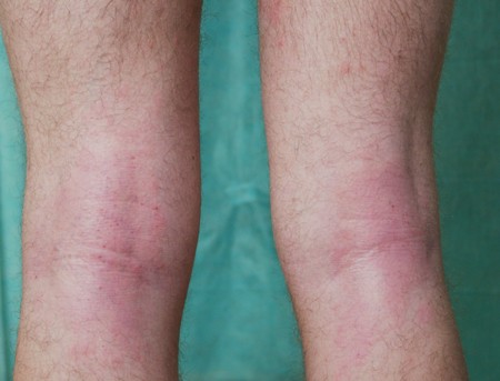
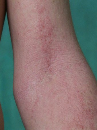
Dermatitis herpetiformis
History
history of gluten intolerance; presence of other autoimmune diseases; exacerbation of symptoms due to iodide exposure (e.g., after eating shellfish)
Exam
presence of pruritic erythematous papules, tiny vesicles, and small blisters over knees, elbows, shoulders, scalp, and buttocks
1st investigation
- direct immunofluorescence of skin biopsy:
granular deposits of IgA in the dermal papillae
- detection of antibodies to gliadin, reticulin, endomysium, and tissue transglutaminase (mainly IgA):
presence of antibodies against smooth muscle endomysium, gliadin, transglutaminase, reticulin
Other investigations
- histology of skin biopsy:
presence of subepidermal blisters that have papillary micro-abscesses at their periphery
Bullous pemphigoid
History
sometimes a history of drug intake (e.g., ACE inhibitors, diuretics); blistering eruptions occur mainly in older people
Exam
1st investigation
- direct immunofluorescence of skin biopsy:
deposits of IgG and C3 along the dermo-epidermal junction
More
Other investigations
- indirect immunofluorescence:
detection of circulating autoantibodies of pemphigoid type (anti-BP1 and anti-BP2)
More - histology:
subepidermal blisters with perivascular infiltrates consisting of lymphocytes and eosinophils in the dermis
- blood smear:
eosinophilia (>400 eosinophils/microliter)
Iron deficiency anemia
History
sometimes a history of bleeding, otherwise nonspecific
Exam
signs in addition to pruritus: glossitis, angular cheilitis, pale mucous membranes
1st investigation
- blood morphology:
MCV low; MCH low; hemoglobin low
Other investigations
- iron serum level:
low
More - total iron binding capacity (TIBC):
elevated
- serum ferritin:
low
Diabetic peripheral neuropathy
History
history of type 1 or type 2 diabetes; risk factors include prolonged duration of diabetes (e.g., >10 years), older age (e.g., >70 years), tall stature, and hyperglycemia; may be asymptomatic; symptoms of peripheral neuropathy include pain, dysesthesias, numbness, weakness is less common; may be associated with features of autonomic neuropathy (orthostatic hypotension, gastroparesis, esophageal dysfunction, diarrhea, fecal incontinence, bladder dysfunction, and erectile dysfunction)
Exam
loss of vibration sense and light touch (may show stocking and glove distribution), reduced ankle reflexes, injuries without pain (especially feet)
1st investigation
- none:
clinical diagnosis
Other investigations
- nerve conduction tests:
reduced sensory nerve conduction velocity and decreased amplitude
More - electromyogram (EMG):
may be normal in mild or asymptomatic patients or show denervation in more severe neuropathy
- quantitative sensory testing (QST):
may be normal or show reduced vibration and/or thermal perception threshold
Hodgkin lymphoma
History
prolonged generalized pruritus that often precedes diagnosis
Exam
enlargement of peripheral lymph nodes, fever, malaise, skin xerosis
1st investigation
- biopsy of lymph nodes:
histologic features of Hodgkin disease
Other investigations
- sonography:
hepatomegaly, splenomegaly, enlargement of lymph nodes
Polycythemia vera
History
venous or arterial thrombosis; pricking-type itch persisting for hours after a hot shower or bath;[64][65] some patients may also have aquagenic pruritus (i.e., intense itching sensation that develops immediately after contact with water at any temperature), headache, dizziness, and acral paresthesias[66]
Exam
signs of thrombosis (e.g., cyanotic face skin color, often plethoric with blue-red tone; often, darkened oral mucous membranes)
1st investigation
- blood morphology:
erythrocytes elevated; leukocytes elevated; platelets elevated; anisocytosis and poikilocytosis of erythrocytes; hemoglobin elevated; hematocrit elevated
Other investigations
- sonography:
hepatomegaly, splenomegaly
- bone marrow aspirate:
increased cell density with numerous erythroblasts and megakaryocytes
HIV infection/AIDS
History
history of drug misuse (especially intravenous); multiple sexual partners; travelers from endemic regions, especially those with a history of transfusion or unprotected sexual intercourse
Exam
features typical of one of the commonly associated dermatologic conditions such as HIV-associated eosinophilic folliculitis, pruritic papular eruption, xerosis, seborrheic dermatitis, psoriasis, scabies, superficial fungal infections, drug eruptions, urticaria, photosensitivity reactions
1st investigation
- serologic tests:
identification of antibodies against HIV components
More
Thyroid dysfunction
History
generalized pruritus develops in 4% to 11% of patients with hyperthyroidism;[67] pruritus also occurs with hypothyroidism, mainly due to skin xerosis; symptoms of hypothyroidism include weight gain, cold intolerance, fatigue, constipation, dry skin; symptoms of hyperthyroidism include anxiety, heat intolerance, palpitations, weight loss, weakness, eye problems, diarrhea, tremor, moist skin
Exam
hypothyroidism: bradycardia, waxy and dry skin, myxedema; hyperthyroidism: tachycardia, thyroid enlargement, tremor, exophthalmos, moist and smooth skin
1st investigation
- thyroid-stimulating hormone (TSH):
low (hyperthyroidism) or elevated (hypothyroidism)
More
Other investigations
- fT4:
low (hypothyroidism) or elevated (hyperthyroidism)
- fT3:
low (hypothyroidism) or elevated (hyperthyroidism)
Paraneoplastic pruritus
History
generalized pruritus may be the first and only symptom of various malignancies (e.g., carcinoid; cancers of the breast, stomach, lungs, vulva, prostate, colon, rectum; leukemias; lymphomas); symptoms of underlying malignancy may also be present; sometimes pruritus may occur in the late stages of cancer disease accompanying cachexia and requiring palliative treatment[68]
Exam
generalized pruritus, sometimes associated with xerosis
1st investigation
- imaging (sonography, x-ray, CT, MRI, scintigraphy, and others, depending on suspected malignancy):
presence of various tumors
More
Other investigations
- blood morphology:
frequently anemia: hemoglobin low; leukocytosis: leukocytes elevated
More - erythrocyte sedimentation rate (ESR):
>20 mm after 1 hour
More - C-reactive protein (CRP):
elevated
More - biopsy:
malignant cells
More - other (depending on suspected malignancies):
various tests can demonstrate abnormalities during the course of malignant disease, depending on the type of cancer
More
Brachioradial pruritus
History
history of symmetric pruritus in the elbow region, over the proximal muscle-tendon attachments of brachioradial muscle; pruritus sometimes extends across the back and occasionally to the chest; patients frequently report exacerbation after sun exposure[48]
Exam
usually no visible abnormalities of skin; secondary changes (e.g., lichenification and/or hyperpigmentation) due to chronic rubbing or scratching may sometimes be present
1st investigation
- cervical spine x-ray:
abnormalities in C5-C8 region
More
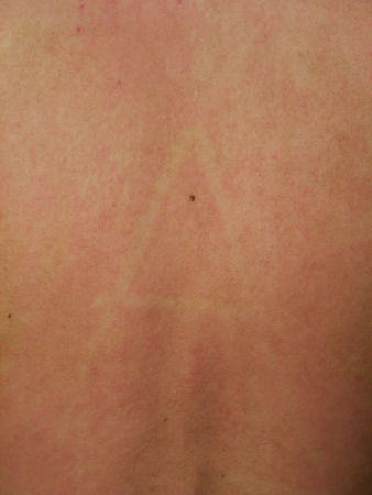
Notalgia paresthetica
History
localized, usually unilateral itching on back near scapula in T2-T6 dermatomes; pruritus may be accompanied by other symptoms such as burning, tingling, numbness, or formication; notalgia paresthetica may also be a neurologic symptom in patients with multiple endocrine neoplasia 2A (medullary thyroid carcinoma, pheochromocytoma, parathyroid hyperplasia) that must be excluded[49][70]
Exam
usually no visible skin abnormalities, sometimes secondary changes (e.g., lichenification and/or hyperpigmentation) due to chronic rubbing or scratching may be present
1st investigation
- thoracic spine x-ray:
abnormalities in the T2-T6 region
More
Brain tumor
History
localized pruritus (e.g., affecting nose and face); patient may present with other neurologic symptoms/deficits indicating brain tumor: for example, symptoms of raised intracranial pressure (headache, altered mental status, nausea and/or vomiting)
Exam
focal neurologic deficits according to tumor location; gait abnormality; papilledema in raised intracranial pressure
1st investigation
- MRI head:
presence of tumor in the brain
More
Other investigations
- CT head:
presence of tumor in the brain
More
Stroke
History
history of unilateral neurogenic pruritus; other more common symptoms of stroke include limb and/or facial weakness, paresthesias or numbness, speech difficulty, headache, visual loss or double vision, confusion, dizziness, vertigo, nausea, neck or facial pain, impaired coordination
Exam
focal neurologic deficits according to site of stroke may include hemiplegia, motor hemiparesis, sensory hemiparesis, dysphasia or aphasia, dysarthria, ataxia
1st investigation
- CT:
presence of ischemic/hemorrhagic area in the brain
More
Other investigations
- MRI:
presence of ischemic/hemorrhagic area in the brain
More
Multiple sclerosis
History
paroxysmal, symmetric, segmental itching that can last from several seconds to few minutes; itching attacks that often awaken patient from sleep; episodes may be spontaneous or triggered by a bath or sudden movement[26]
Exam
witnessed itching and excoriations may be present in any part of the body
1st investigation
- MRI of brain and spine:
bright spots on image that correspond to areas of demyelination
More
Other investigations
- cerebrospinal fluid:
oligoclonal band in protein electrophoresis, evidence of chronic inflammation
More
Psychogenic pruritus
History
compulsory criteria: localized or generalized, chronic (>6 weeks) pruritus without primary skin lesions and other somatic cause; additional criteria (3 of 7 have to be present): chronologic relationship of pruritus with 1 or several life events that could have psychological repercussions, variations in the intensity associated with stress, nocturnal variations, predominance during rest or inaction, associated psychological disorders, pruritus that could be improved by psychotropic drugs, pruritus that could be improved by psychotherapies[72]
Exam
no physical signs of skin diseases and other disorders that may be responsible for pruritus
1st investigation
- none:
diagnosis is clinical
More
Other investigations
Persistent delusional disorders
Filariasis
History
urticaria, pruritus, migrating worm may be seen under the skin
Exam
dermatitis and nodules; Loa loa infestation more likely to present with visual defects and signs of eye infection
1st investigation
- CBC:
eosinophilia
- Giemsa-stained blood smear:
identifies microfilariae
- peripheral blood smear:
identifies microfilariae
Other investigations
Strongyloides infection
History
history of migration from endemic part of world; abdominal pain, altered bowel habit, and weight loss are common symptoms; patient may complain of pruritus
Exam
dermatitis, larva currens; if hyperinfection there may be signs of sepsis
1st investigation
- stool examination:
ova and parasites
- CBC:
eosinophilia
Other investigations
- chest x-ray:
pulmonary infiltrates
- skin or tissue biopsy:
strongyloides larvae
Use of this content is subject to our disclaimer