Tests
1st tests to order
knee radiographs
Test
Views should include a standing anteroposterior view of the bilateral knees, a lateral view at 30º of flexion, a merchant or skyline view of the patellofemoral joint, and a notch or posteroanterior (PA) flexion view. Particular attention should be paid on the posterolateral aspect of the medial femoral condyle, which is often best seen on the notch or PA flexion view.
Result
osteochondral lesion, free intra-articular loose bodies, malalignment, or arthrosis
ankle radiographs
Test
Views should include anteroposterior, lateral, and mortise of the ankle.
Result
osteochondral lesion, free intra-articular loose bodies, malalignment, or arthrosis
full-length lower extremity film
Test
Lower extremity alignment should be assessed with a full-length lower extremity film. If malalignment exists and the weight-bearing line passes through the involved compartment, some orthopedic surgeons will consider an osteotomy to unload the involved compartment.[3]
Result
may show malalignment with weight-bearing line passing through involved compartment
elbow radiographs
Test
Views should include anteroposterior, lateral, and internal and external oblique.
Result
osteochondral lesion, free intra-articular loose bodies, malalignment, or arthrosis
Tests to consider
CT
Test
CT is very useful in identifying loose bodies within the joint. The sharp contrast between bone and soft tissue, along with the ability to obtain fine slices, make CT a good tool to identify loose bodies trapped within joint recesses and synovial folds. The ability to obtain axial, coronal, and sagittal images further enhances its utility in identifying osteochondral injury. Do not order CT scans for children until plain radiographic examinations have been completed.[24]
Result
may identify loose bodies within the joint and trapped within joint recesses and synovial folds; osteochondral injury
MRI
Test
MRI allows direct visualization of the chondral surface in axial, coronal, and sagittal planes. MRI has been shown to be a useful noninvasive mechanism to identify osteochondral lesions.[25][26][27] It helps delineate the stability of the lesion as well as the location and number of lesions and loose bodies. The sensitivity of MRI to determine the extent of chondral injury, compared with arthroscopy, has ranged from 33% to 96% in available studies and usually underestimates the extent of the lesion.[28] MRI imaging protocols for juvenile osteochondritis dissecans are often center-specific. Ideally, protocols should include a cartilage-sensitive sequence and a fluid-sensitive sequence. Do not order MRI scans for children until plain radiographic examinations have been completed.[24] Attention is given to the amount of localized marrow edema, as well as lesion location and size, and the stability of an osteochondral fragment.[Figure caption and citation for the preceding image starts]: Coronal magnetic resonance image (MRI) of the talus showing an osteochondral lesion on the medial aspect of the talar domeGupta RK, Kansay R, Aggarwal V, et al. Osteochondritis dessicans of the talus in a 26-year-old woman. BMJ Case Reports 2009; doi:10.1136/bcr.06.2008.0091 [Citation ends]. [Figure caption and citation for the preceding image starts]: Sagittal magnetic resonance image (MRI) of the talus, showing an osteochondral lesion on the posterior aspect of the talar domeGupta RK, Kansay R, Aggarwal V, et al. Osteochondritis dessicans of the talus in a 26-year-old woman. BMJ Case Reports 2009; doi:10.1136/bcr.06.2008.0091 [Citation ends].
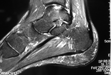
Result
osteochondral lesions; loose bodies
MR arthrogram
Test
Can provide additional information regarding the articular surface. Intra-articular administration of contrast can be performed in the absence of a large joint effusion. This can aid the accuracy of MRI to diagnose subtle chondral defects and to determine the stability of the osteochondral lesion. Intravenous administration of gadolinium, which is then secreted by the synovium, improves visualization of bony edema and osteochondral lesions.[32][33] MR arthrogram is particularly helpful in the elbow, as cartilaginous loose bodies can be identified.
Result
subtle chondral defects; stability of the osteochondral lesion; with intravenous gadolinium administration, shows bony edema and osteochondral lesions
diagnostic arthroscopy
Test
This remains the most specific and sensitive test, but as an invasive procedure, its role in initial diagnosis should be limited with the advanced imaging techniques available today. If performed it allows other diagnoses to be ruled out.
Result
direct visualization of the osteochondral lesion
Use of this content is subject to our disclaimer